
Dr Andrew Baron
Scientific Director of Biosimilars, Product Characterisation and Biophysics

 Insights
|
November 5, 2026
|
8 min read
Fc effector function is the set of immune activities an antibody triggers through its Fc region rather than through the arms that bind its target. Whether your candidate biologic needs those activities retained, enhanced or removed, you can determine which it has by measuring binding to the Fcγ receptors and C1q, then confirming what that binding does in cell-based assays.
This guide covers what Fc effector function is, how to assess it, how Fc receptor binding is measured by surface plasmon resonance, how to test an Fc-engineered antibody, which ADCC format to choose, how ADCP and complement activation are measured, how to confirm Fc silencing and a published example of PBMC-based ADCC data in a biosimilar comparability package.
Fc effector function is the activity an antibody drives through its Fc region by engagement of Fc gamma receptors (FcγRs) on immune cells such as natural killer (NK) cells, macrophages and neutrophils, and binding of the complement protein C1q. The three functions most often measured are antibody-dependent cellular cytotoxicity (ADCC), antibody-dependent cellular phagocytosis (ADCP) and complement-dependent cytotoxicity (CDC).
It is essential because effector function can be the mechanism, the liability or the comparability attribute. In some therapeutic antibodies, Fc effector function contributes directly to efficacy by eradicating target cells. In others, such as many checkpoint inhibitors and receptor-blocking antibodies, it may be reduced or eliminated to avoid unwanted immune activation. For a biosimilar, it is an attribute that has to match the reference product, often whether it is present or absent.
The key consideration is that Fc effector function should be appropriate for the intended mechanism of action, whether that means retaining, reducing or enhancing activity. That applies across monoclonal antibodies, bispecifics, multi-specifics, immune cell engagers, antibody-drug conjugates (ADCs) and Fc-fusion molecules, because each carries an Fc region whose behaviour has to be characterised.
The neonatal Fc receptor (FcRn) is a separate question. It governs recycling and half-life rather than effector function, and we cover it in our guides to the role of FcRn in immunology and therapeutics and FcRn-mediated recycling assays.
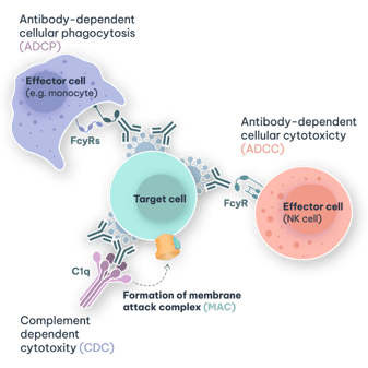
Figure 1. Key Fc-mediated effector functions of monoclonal antibodies, including ADCC, ADCP and CDC, driven through interactions with Fcγ receptors or complement.
You assess it in two layers: measure binding to the Fcγ receptors and C1q, then confirm what that binding produces in cell-based functional assays with the relevant effector cells or complement source. Binding data demonstrates what the Fc is capable of engaging, and functional data demonstrates what happens when it does.
Both layers are needed because the relationship between them is not fixed. In our own IgG variant panel, an IgG1 LALA variant retained 42% of wild-type FcγRIIIa V binding yet mediated 87% of wild-type ADCC, so binding alone would have under-estimated the residual function (Kerr, 2026). The reverse also happens: trastuzumab binds C1q but does not mediate CDC (Metikala et al., 2026).
| Endpoint | What it measures | Typical method | Potential functional relevance |
|---|---|---|---|
| FcγRI (CD64) binding | Engagement of the high-affinity activating receptor. | SPR or BLI. | ADCP. |
| FcγRIIa (CD32a, H and R variants) binding | Engagement of the low-affinity activating receptor on myeloid cells. | SPR or BLI. | ADCP. |
| FcγRIIb (CD32b) binding | Engagement of the inhibitory receptor. | SPR or BLI. | Immune regulation, and crosslinking for some agonist antibodies. |
| FcγRIIIa (CD16a, V and F variants) binding | Engagement of the activating receptor on NK cells. | SPR or BLI. | ADCC. |
| FcγRIIIb (CD16b) binding | Engagement of the neutrophil receptor. | SPR or BLI. | Neutrophil involvement. |
| C1q binding | The first step of classical complement activation. | SPR or BLI. | CDC potential. |
| FcRn binding | pH-dependent recycling. | SPR. | Half-life, not effector function. |
| ADCC | Killing of target cells by FcγRIIIa-bearing effector cells. | Reporter cell line, or primary NK cells or PBMCs. | Mechanism, potency, comparability. |
| ADCP | Phagocytosis of target cells by FcγRI/FcγRIIa-bearing cells. | Reporter cell line, or monocyte-derived macrophages. | Mechanism, potency, comparability. |
| CDC | Complement-mediated lysis of target cells. | Target cells with a complement source. | Mechanism, potency, comparability. |
The FcγRIIIa polymorphism deserves a note on numbering. The higher-affinity valine and lower-affinity phenylalanine alleles are written V158 and F158 in most of the literature, and V176 and F176 where the numbering includes the signal peptide. Both alleles are usually tested, and this guide uses 158 except where it quotes a paper that uses 176.
You capture the one binding partner to the sensor surface as the ligand and flow over a concentration series of the other as the analyte, so association and dissociation are measured in real time and an affinity (KD) is derived for each interaction.
Capture orientation. In the design we qualified for an anti-HER2 IgG1, the antibody is captured through the light chain in the Fab region and the receptors flow over it in as the analyte. That reduces avidity effects and removes the influence of product formulation on the measurement (Moncrieff and Julien, 2026). We run these assays on Biacore 8K systems using Fc receptors produced in-house.
Reagents. Our recombinant Fc receptor panel covers eight human receptors (FcγRI, FcγRIIa H and R variants, FcγRIIb, FcγRIIIa V and F variants, FcγRIIIb and FcRn), five cynomolgus receptors and five murine receptors. They are expressed in HEK293 cells, C-terminally histidine tagged, purified to above 95% by SDS-PAGE and checked for bioactivity by SPR.
The cynomolgus set allows species relevance to be assessed at the Fc as well as at the target, which is covered in our guide to the in vitro data package from lead selection to IND.
Reporting. Relative affinity, expressed as a percentage of a reference standard tested in the same run, holds up better from day to day than absolute KD, ka and kd values. Relative values are therefore what typically go into a comparability package, with the absolute values retained for characterisation.
Qualification. For regulatory use the methods are qualified against ICH Q2(R2). In the anti-HER2 study, eight SPR methods (seven Fcγ receptor binding assays plus FcRn and HER2 binding) met pass criteria of 80 to 125% for accuracy, a coefficient of variation of 20% or below for repeatability and intermediate precision, and a specificity control response of 20% or below of the reference material.
The specificity control for the Fcγ receptor assays was an IgG1 carrying the L234S, L235T and G236R substitutions, which abolish Fcγ receptor binding (Wilkinson et al., 2021).
Reagent quality decides how far any of this can be trusted, so it is worth reading our guide to key materials and reagents for SPR before designing the study.
Match the assay panel to the design. An Fc engineered to enhance ADCC should be characterised for the gain it was built for, and an Fc engineered to be silent should be characterised for what remains, in both cases with binding and function measured side by side.
Three designs cover most engineered antibodies:
Our own data demonstrates why the silenced designs need measuring rather than assuming. In a panel of IgG1 wild type, IgG2, IgG4, LALA, LALAPG and STR variants tested in SPR binding and primary cell functional assays, IgG1 wild type demonstrated strong FcγRIIIa binding with ADCC activity, and high C1q binding with CDC activity. Most of the variant panel significantly reduced these functions.
The LALA variant, which is typically referred to as silenced, retained 42% of wild-type FcγRIIIa V binding and mediated 87% of wild-type ADCC activity. LALA, IgG2 and IgG4 also retained binding to CD64 and CD32, the receptors most relevant to ADCP. In this panel, only the STR substitutions abolished Fc receptor binding and effector function while preserving other Fc properties (Kerr, 2026; Miller, 2024).
Fc silencing is therefore a matter of degree. The clinical significance of any residual activity should be considered carefully for the format in question, which is only possible once it has been measured.
Neither is best in the abstract, because they answer different questions. A reporter assay gives a reproducible potency readout suited to comparability and lot release, while primary effector cells report the killing of target cells by human immune cells and are the more physiologically relevant read.
Reporter cell lines. Jurkat cells engineered to express FcγRIIIa, in the V or F variant, with a luciferase gene under the control of nuclear factor of activated T cells (NFAT) response elements. The signal reports receptor engagement and signalling rather than killing. Variability is low, there is no donor to source and the format lends itself to qualified potency methods.
Primary NK cells or PBMCs. Peripheral blood mononuclear cells (PBMCs) or isolated NK cells from donors genotyped for FcγRIIIa. The readout is loss of target cells, which is the biological event the assay is meant to predict. Variability is higher and donor-dependent, which is why controlled donor material and genotyped, recallable donors count over the life of a programme.
| Reporter cell line | Primary NK cells or PBMCs | |
|---|---|---|
| What the readout reports | Fc receptor engagement and signalling in an engineered cell. | Killing of target cells by human effector cells. |
| Variability | Low. | Higher, and donor-dependent. |
| Donor genotype | Fixed by the engineered line, V or F. | Selected by genotyping donors, and both alleles can be tested. |
| Throughput | High. | Lower. |
| Best use | Characterisation, lot release and screening. | Mechanism elucidation, characterisation and confirming absence of function. |
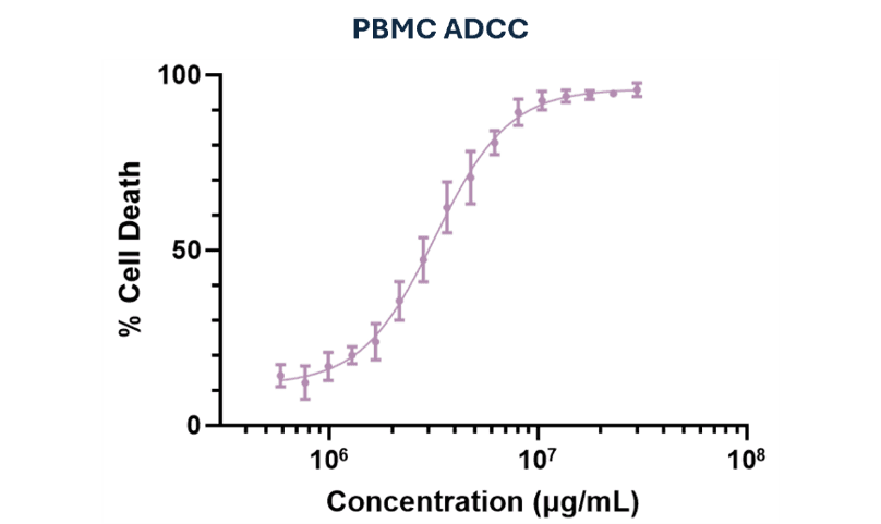
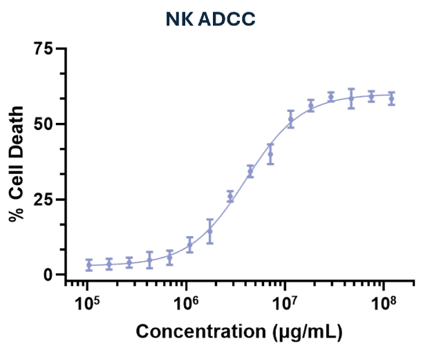
Figure 2. ADCC activity measured using NK cell- and PBMC-based assay formats, illustrating concentration-dependent target-cell killing across different effector cell models.
Many packages use both, and the published biosimilar study later in this guide ran V and F reporter assays alongside PBMC-based assays with FF and VV donors.
The target cells are important just as much as the effectors. An engineered line expressing the antigen at a defined density, ideally carrying a luciferase or other constitutive readout, gives a consistent target, and the density you choose can change the result, as our guide to target antigen density explains.
We build target lines in-house through our custom cell engineering service, and RoukenCells sells stable expression cell lines and reporter cell lines off the shelf.
Readouts include luciferase, flow cytometry and real-time impedance.
ADCP is measured by presenting antibody-coated target cells to FcγRIIa-bearing effector cells and reading either phagocytosis or receptor signalling. The relevant receptors are FcγRIIa (CD32a), with contributions from FcγRI (CD64), on monocytes and macrophages.
In the primary format, monocyte-derived macrophages are co-cultured with labelled target cells and phagocytosis is quantified by flow cytometry or imaging. In the reporter format, Jurkat cells expressing FcγRIIa with an NFAT-driven luciferase report engagement in the same way as the ADCC reporter line.
ADCP is worth testing directly rather. In our variant panel, LALA, IgG2 and IgG4 retained binding to CD64 and CD32, so ADCP is the effector function may persist after a silencing strategy aimed at FcγRIIIa. Our macrophage assays page and our technical presentation on macrophages in immunity and immuno-oncology cover the primary formats in more detail.
You assess it in two steps: C1q binding by SPR, ELISA or bio-layer interferometry (BLI), then complement-dependent cytotoxicity (CDC) in antigen-expressing target cells with a complement source, read as lysis. In the SPR binding assay, the antibody is captured and C1q is flowed over it; in the functional assay the antibody, target cells and complement are combined and the loss of target cells is measured.
Binding does not equal function here either. Trastuzumab binds C1q but has no CDC activity, and the biosimilar study described below confirmed that absence with a positive control (Metikala et al., 2026).
That control, rituximab on CD20-expressing WIL2-S cells, gave 1.11 to 2.53-fold activity over the complement-only control across its dose range, while no CDC was observed for trastuzumab at any dose. A negative CDC result only means something alongside a positive control run in the same assay.
In our own variant panel, IgG1 wild type showed high C1q binding and CDC activity, while the variants showed minimal complement activity overall, with only modest residual activity for IgG1 LALA. Our bioanalytical assessments page sets out the Fcγ and C1q binding and effector function assays we run.
Absence of effector function takes more evidence to demonstrate than presence, because a negative result can come from a silent Fc or from an assay that could not have detected activity. Positive in-assay controls, sensitive functional formats and orthogonal binding data are what tell the two apart.
For a biosimilar in an IgG2, IgG4 or engineered IgG1 format, regulators including the CDSCO, FDA and EMA look for robust evidence that pertinent biological activities, present or absent, are matched to the reference product, because differences may affect clinical outcomes. Our biosimilars page describes how these assays fit a comparability package.
The same logic applies to a novel silenced biologic. The question is rarely whether a silencing strategy was used, but what residual activity remains and whether it is likely to make a difference in the clinic.
In September 2026, CuraTeQ Biologics published a comparative analytical assessment of BP02, its trastuzumab biosimilar, against EU-approved and US-licensed Herceptin in Scientific Reports (Metikala et al., 2026). We developed the PBMC-based ADCC methodology used in that study and carried out the sample testing, and the resulting data appears as Figure 13 G and H of the paper.
The study measured ADCC two ways. Reporter gene assays used engineered Jurkat cells expressing FcγRIIIa V176 or F176 with BT474 target cells. The PBMC-based assay used HER2-expressing CHO-K1 luciferase target cells with PBMCs from FF and VV donors tested separately, read by luciferase signal, with relative potency calculated against the reference standard.
In the PBMC-based assay, BP02 gave a mean relative potency of 105% with VV donors and 102% with FF donors across seven batches, within the Herceptin quality ranges, which the authors set at the reference product mean plus or minus three standard deviations. The reporter gene assays reached the same conclusion, and the paper reports that BP02 and Herceptin were similar in inducing ADCC.
Two further findings in the paper illustrate points made above. One BP02 batch fell outside the Herceptin range for FcγRIIIa F176 binding by SPR yet gave in-range ADCC potency in both the reporter and PBMC-based assays. In this case the functional assay settled the question.
The paper's acknowledgement reads: "We thank RoukenBio, Scotland for their support in developing the PBMC-based ADCC methodology and sample testing." The full paper is open access.
Many antibody developers run ADCC, ADCP and CDC assays in-house. External support becomes particularly valuable when a programme needs assay formats, reagents or capabilities that are not routinely available internally, or when additional sensitivity, physiological relevance or assay development is required.
At RoukenBio, we combine FcγR and C1q binding by SPR with functional ADCC, ADCP and CDC assays. Depending on the biological question, these can incorporate engineered target cells, genotyped, functionality-screened and recallable primary-cell donors, or reporter formats.
This can be especially useful when investigating residual activity in an Fc-silenced molecule, comparing different Fc designs, understanding an unexpected functional result, or developing an assay towards a defined regulatory purpose.
Because effector function is rarely the whole biological story, we start with the mechanism and the question that needs to be answered, rather than a fixed assay list. The resulting strategy can then reflect the molecule, its intended mechanism of action and its stage of development.
View our Bisomilars Capabilities
Fc effector function is the set of immune activities a therapeutic antibody triggers through its Fc region rather than through its antigen-binding arms. The Fc region engages Fc gamma receptors on natural killer cells, macrophages and neutrophils, and binds the complement protein C1q. The resulting functions are antibody-dependent cellular cytotoxicity, antibody-dependent cellular phagocytosis and complement-dependent cytotoxicity.
Get an in-depth look at our biosimilar testing approach through our detailed slide deck, featuring sample data and a real-world biosimilar case study.

🗓️ Stay informed with our monthly scientific newsletter, published on LinkedIn on the last Wednesday of each month.
These editions bring you the latest in drug development breakthroughs, industry trends, and expert insights from the brilliant minds at RoukenBio.
Subscribe today on LinkedIn
 Research Posters
 Webinars