We offer a comprehensive suite of macrophage assays to support your immunological research and drug development projects. Our ability to differentiate monocyte-derived macrophages (MDM) into M1 and M2 populations for use in a variety of assayWe offer a comprehensive suite of macrophage assays to support your immunological research and drug development projects. Our ability to differentiate monocyte-derived macrophages (MDM) into M1 and M2 populations for use in a variety of assays, including macrophage polarisation, macrophage phagocytosis readouts, and antibody-driven ADCP assays, ensures you can evaluate your immunotherapies in an appropriate disease context.

Because macrophages can adopt pro- or anti-inflammatory states depending on local cues. Well-designed macrophage functional assays are essential for understanding whether a therapeutic enhances or blocks inflammatory activation, reprogrammes suppressive phenotypes, or modifies clearance mechanisms such as phagocytosis.
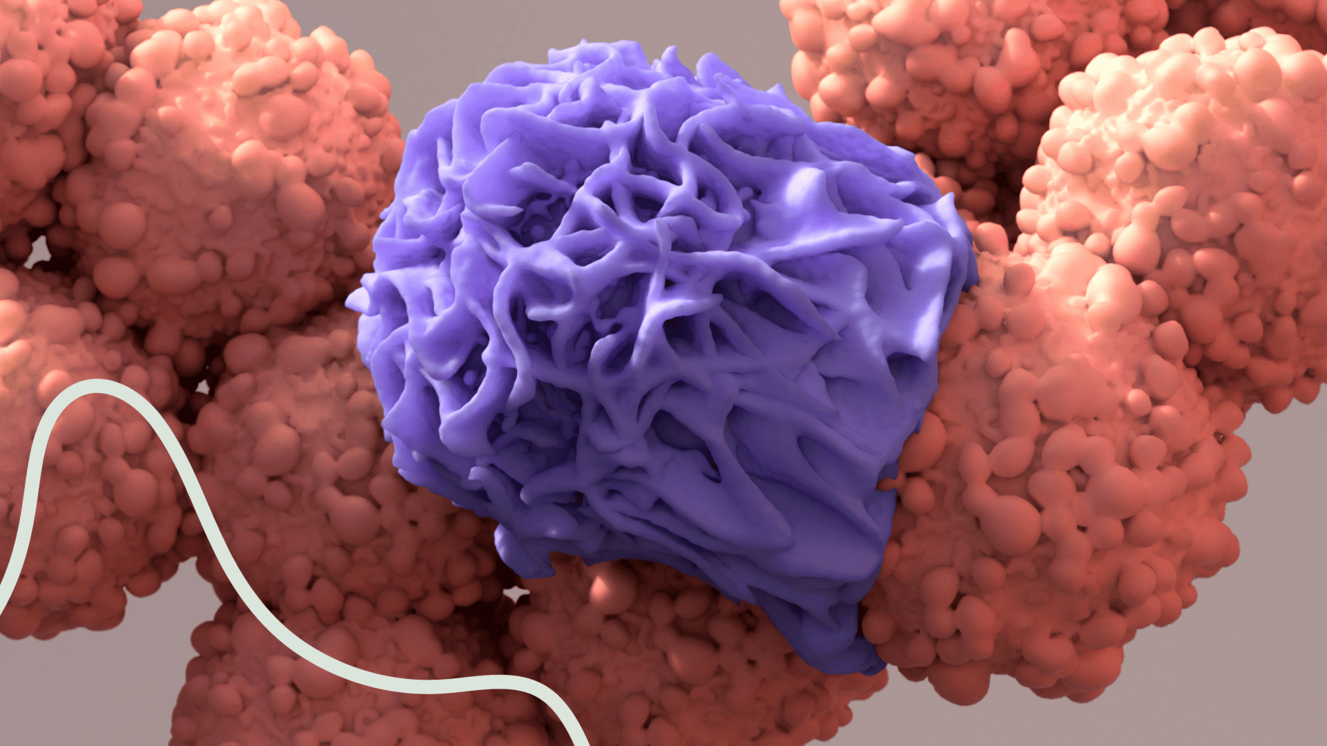
Macrophages are specialised cells of the innate immune system performing a range of functions including the detection and destruction of pathogens, initiation and resolution of the immune response, immunosuppression, waste disposal, wound healing and angiogenesis. These cells can be anti-tumourigenic (cancer cell phagocytosis and antigen presentation) or pro-tumourigenic (immune suppression and growth factor secretion). Similarly, macrophages can also have opposing roles in the development and control of autoimmune and fibrotic diseases.
Macrophage polarisation
Macrophage Mediated ADCP
Phagocytosis
Macrophage Activation of T cells
Our macrophage polarisation assays help assess how candidate therapeutics modulate macrophage phenotype, determining whether they promote pro-inflammatory activation or drive differentiation towards an immunosuppressive state.
We generate monocyte-derived macrophages (MDMs) that can be polarised in a variety of ways to reflect the disease indication most relevant to your research. CD14+ monocytes are differentiated using M-CSF to generate resting state (M0) macrophages. These can then be polarised and matured using a variety of stimuli to reflect different sub-populations e.g., M1, M2a, M2c, M2d – associated with particular pathological processes, e.g. M2 tumour associated macrophages (TAM). Our assays allow you to determine the impact of your candidate therapeutic on blocking or even reprogramming macrophage profiles, assessing phenotypic and functional changes to reveal switches in pro- and anti-inflammatory responses.
Identifying MDM by Flow Cytometry:

Macrophages are easily identified by CD64+CD11b+ phenotype.
Distinguishing Sub-types:

Polarisation of monocytes into specific macrophage sub-populations can by analysed by Flow Cytometry. Test samples can be assessed for their ability to impact M0 polarisation to particular sub-populations or assessed for their ability to switch or alter existing macrophage phenotype.
TNF∝ Secretion by MDM Sub-types:

Macrophages can be assessed for cytokine production by ELISA or multiplex cytokine analysis. Typically, M1 are potent producers of TNF∝, consistent with pro-inflammation. M2 are poor sources of TNF∝.
Our macrophage polarisation assays help assess how candidate therapeutics modulate macrophage phenotype, determining whether they promote pro-inflammatory activation or drive differentiation towards an immunosuppressive state.
We generate monocyte-derived macrophages (MDMs) that can be polarised in a variety of ways to reflect the disease indication most relevant to your research. CD14+ monocytes are differentiated using M-CSF to generate resting state (M0) macrophages. These can then be polarised and matured using a variety of stimuli to reflect different sub-populations e.g., M1, M2a, M2c, M2d – associated with particular pathological processes, e.g. M2 tumour associated macrophages (TAM). Our assays allow you to determine the impact of your candidate therapeutic on blocking or even reprogramming macrophage profiles, assessing phenotypic and functional changes to reveal switches in pro- and anti-inflammatory responses.
Identifying MDM by Flow Cytometry:
Macrophages are easily identified by CD64+CD11b+ phenotype.
Distinguishing Sub-types:
Polarisation of monocytes into specific macrophage sub-populations can by analysed by Flow Cytometry. Test samples can be assessed for their ability to impact M0 polarisation to particular sub-populations or assessed for their ability to switch or alter existing macrophage phenotype.
TNF∝ Secretion by MDM Sub-types:
Macrophages can be assessed for cytokine production by ELISA or multiplex cytokine analysis. Typically, M1 are potent producers of TNF∝, consistent with pro-inflammation. M2 are poor sources of TNF∝.
An ADCP assay quantifies macrophage-mediated phagocytosis of antibody-coated targets applicable to I-O studies, therapeutic antibody development and infectious disease research.
RoukenBio employs custom assay where we can guide the selection of an appropriate target cell to assess the ability of your therapeutic antibody to directly drive ADCP or utilise one of our platform ADCP assays to assess indirect effects on ADCP.
A macrophage phagocytosis assay gives you the capability to assess the impact of your therapeutic candidate on the engulfment and digestion of pathogens, particles or cells by macrophages, applicable to infectious disease research and immune response studies alike.
Assessing Phagocytic ability of MDM:

Dose-dependent increase in ADCP with increasing Rituximab concentration. Target cells can be substituted with the relevant target-expressing cell line to assess effector function of other antibodies. A large number of immortalised cell lines are ready for use, as well as our ability to develop target specific cell lines.
A macrophage T cell activation assay evaluates macrophage function via their ability to stimulate T cell responses. Using macrophages to stimulate allogeneic T cells in a one-way MLR allows assessment of their wider ability to support immune responses, driving either pro- or anti- inflammatory adaptive responses. Link macrophage phenotypic data such as expression of co-stimulatory/inhibitory molecules to cytokine production in response to allogeneic lymphocytes. This is applicable to transplant immunology, graft-versus-host disease studies and immunogenicity testing. We have a clear understanding of your needs, wants and challenges – so we can provide the exact support required to achieve your goals.
M1 MDM Drive T cell activation:

M1 MDMs drive robust T cell activation in the MLR with the highest level of T cell proliferation and IFNγ production. 'Anti-inflammatory' M2 MDMs induce a lower level of T cell proliferation and poor IFNγ secretion. Therapeutics intended to drive MDMs towards a more anti-tumour/M1 like phenotype can be tested in this system.
MDM suppression of T cells:

Activated PBMCs are stained with proliferation dye and co-cultured with MDM subtypes from the same donor. M2c MDMs are highly suppressive of T cell proliferation. When compared to M1, M2c elicit a high frequency of suppression, followed by > M0 > M2a > M2d. Test articles can be included within suppression assays to test ability to induce or reduce suppressive capacity across MDM subtypes.
Choose RoukenBio for human primary-cell expertise and mechanism-led macrophage assay design. We deliver decision-grade readouts: polarisation, cytokines, phagocytosis and ADCP; using bespoke target-expressing tools and custom co-cultures, backed by scientists who work as an extension of your team. Ready to move your programme forward?


Macrophage assays are typically performed using monocyte-derived macrophages generated from human peripheral blood. These cells can be differentiated and polarised in vitro to create defined phenotypes, enabling controlled and reproducible assessment of macrophage biology.