As autoimmune disease research has advanced, there has been a marked expansion in immunotherapy strategies designed to selectively suppress or re‑educate pathogenic immune responses while preserving protective immunity. These next‑generation approaches seek to restore immune tolerance through targeted modulation of T cells, B cells, and cytokine pathways, moving beyond broad, non‑specific immunosuppression. At RoukenBio, we bring the expertise and capabilities to screen and evaluate your therapeutics against these emerging targets using physiologically relevant autoimmunity assays.

RoukenBio is an autoimmunity and immunotherapy CRO delivering human-relevant, in vitro autoimmune assays from discovery through IND-enabling stages. We pair translational immunology with advanced primary cell systems to clarify mechanism of action, differentiate candidates, and quantify functional immune modulation.
Our autoimmunity therapeutic development services cover bespoke assay design through execution across key immune pathways, supporting antibodies and antibody-based formats, cytokine/receptor-targeting biologics, and cell-based approaches that modulate pathogenic immunity without broad immunosuppression.
Autoimmune diseases arise when a dysregulated immune system mistakenly targets and attacks the body, leading to chronic inflammation and tissue damage. The complex mechanisms underlying autoimmunity have revealed novel targets for modern therapeutic interventions that modulate the immune response, moving beyond the generalised immunosuppression of traditional therapies.
Modern autoimmunity programmes require bespoke assays that reflect human disease biology and generate interpretable functional readouts. Our portfolio of autoimmune assays includes a range of cell types, established formats and bespoke systems tailored to your mechanism, covering pro-inflammatory pathways driven by effector T cells and pathogenic B cells and regulatory mechanisms mediated by Tregs and macrophages.
These assays support autoimmunity drug discovery by enabling candidate screening, ranking, and characterisation through activation, suppression, phenotype polarisation and immune-cell targeted depletion.
Example assays include:
T cells
Tregs
Macrophages
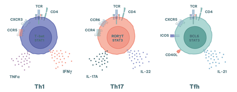
T cells are central to many autoimmune assays because breakdowns in central and peripheral tolerance allow autoreactive T cells to expand, differentiate and drive tissue inflammation. For autoimmunity drug discovery, T cell assays help quantify functional modulation across pathogenic subsets (e.g., Th1, Th17, Tfh) and cytotoxic effector activity.
T cells play a crucial role in the development and progression of autoimmune diseases. In a healthy immune system, T cells help protect the body by directly killing pathogen infected cells and by regulating the immune response. However, in autoimmunity, this system malfunctions, causing T cells to mistakenly target the body's own tissues.
This misdirected attack is often due to a breakdown in the mechanisms that normally ensure self-tolerance, such as thymic selection and peripheral tolerance checkpoints. When these mechanisms fail, self-reactive T cells can proliferate and initiate an immune response against the body's own cells, leading to chronic inflammation and tissue damage.
Different subsets of T cells contribute in different ways to various autoimmune conditions.
They are notably relevant to diseases where neutrophil recruitment is a key pathologic feature. Where the production of high affinity autoantibodies exacerbates disease severity (e.g. SLE and RA), the activity of Tfh cells is also a key consideration.
RoukenBio offers an array of different autoimmunity assay types that can be used to screen drugs and their effects upon T cell differentiation, polarisation and cytotoxicity.
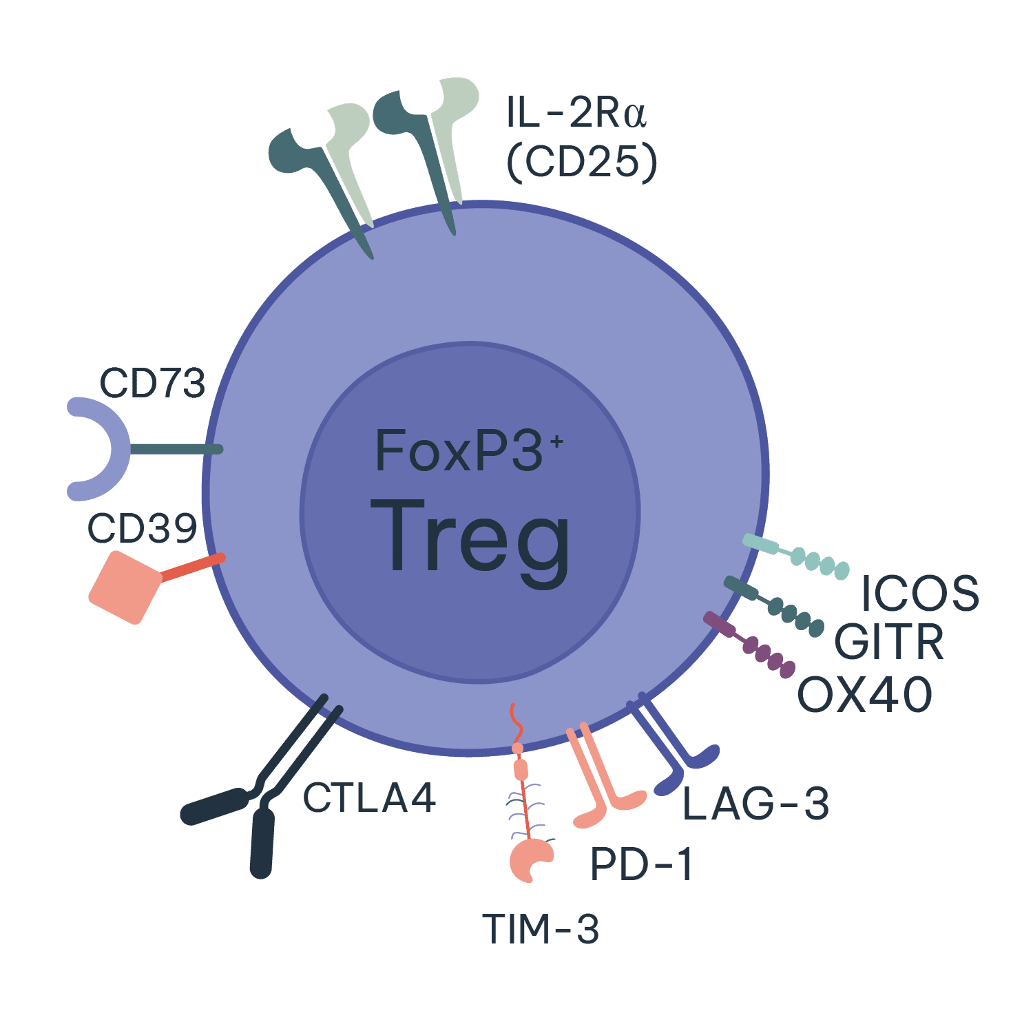
Regulatory T cells (Tregs) maintain immune homeostasis with impaired Treg function implicated in multiple autoimmune diseases. This makes Treg-focused autoimmune assays critical for screening therapies designed to restore immune balance.
In autoimmune diseases, the malfunction or deficiency of Tregs can lead to a loss of self-tolerance, allowing self-reactive T cells to proliferate and cause tissue damage. This dysregulation can result in chronic inflammation and the progression of autoimmune disorders. Enhancing the function or number of Tregs is a promising therapeutic strategy for treating autoimmune diseases, as it can help restore immune balance and reduce pathological immune responses. At RoukenBio, our experts use our Treg induction and suppression assays to help screen and test your Treg targeting therapeutics.
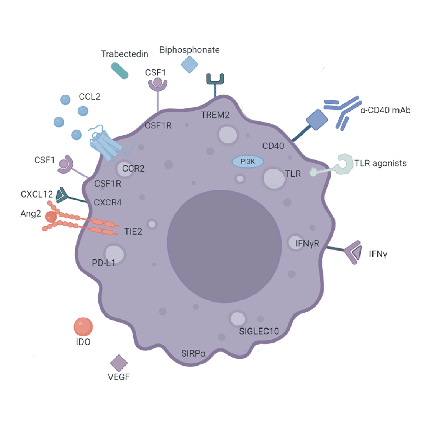
Macrophages shape autoimmune inflammation through antigen presentation, cytokine networks and tissue remodelling, and macrophage state changes (e.g., M1↔M2 polarisation) are commonly assessed using autoimmune assays to understand therapeutic immune reprogramming.
In autoimmunity, macrophages can contribute to disease pathogenesis through their roles in inflammation and tissue damage. They produce various cytokines and chemokines that can either promote or suppress immune responses, depending on the signals they receive from their environment.
In autoimmune diseases, macrophages in the affected tissues exacerbate inflammation and tissue destruction through their role as antigen presenting cells, which induce the activation of autoreactive T cells. In addition, they release pro-inflammatory cytokines like TNF-α and IL-1, which further amplify the immune response and contribute to chronic inflammation.
However, macrophages also have the potential to suppress autoimmune responses and promote tissue repair. Certain subsets of macrophages, known as M2 macrophages, can produce anti-inflammatory cytokines and growth factors that help resolve inflammation and support tissue repair or, in some instances, drive fibrosis.
At RoukenBio, we have a number of macrophage-based assays that can examine macrophage differentiation, activation and suppression.
Mixed Lymphocyte Reaction Assay
Treg Suppression Assays
Macrophage Polarisation Assays
B Cell Depletion Assays
T Cell Depletion Assays
MLR autoimmune assay formats provide a robust, human primary-cell readout of T cell attenuation and immune suppression, supporting early candidate ranking in autoimmunity drug discovery.
Mixed lymphocyte reactions (MLR) are assays in which T cells and antigen presenting cells (APCs) from two genetically mismatched (allogenic) donors are incubated together to induce T cell activation. These assays are commonly used to assess the ability of biologics or small molecules to directly or indirectly alter T cell activation, proliferation and cytokine secretion.
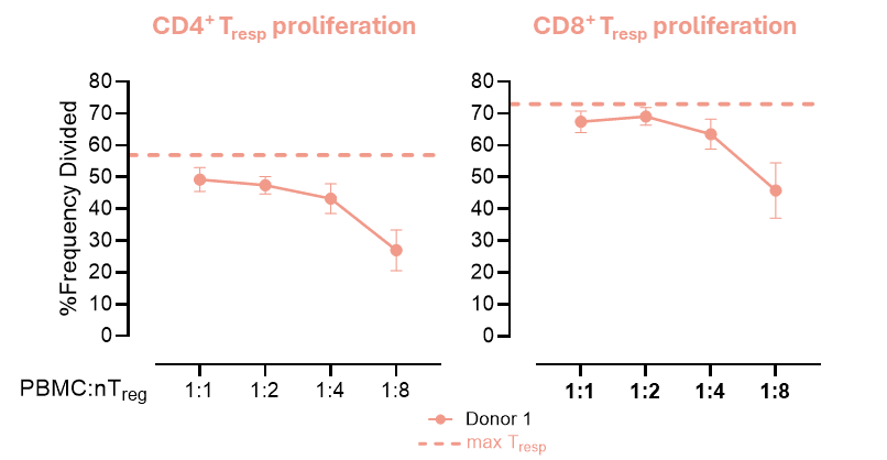
Our autoimmune assays for Treg function quantify suppression of responder T cell proliferation and cytokine release, enabling evaluation of therapeutics designed to induce or enhance immune regulation.
Treg suppression assays are used to evaluate the ability of Tregs to suppress the proliferation and activity of T cell subtypes, such as CD4+ or CD8+ T cells. In these assays, T cells are labelled with a cell tracker dye and incubated with Tregs at different ratios in the presence of a stimulant. The extent to which Tregs inhibit the proliferation and cytokine production of the T cells is then assessed via flow cytometry and ELISA.
These assays are well suited to testing the ability of therapeutics to induce Tregs or to enhance their responses. An example of a Treg suppression assay in which different ratios of Tregs are incubated with stimulated conventional T cells (Tresp) is shown below:
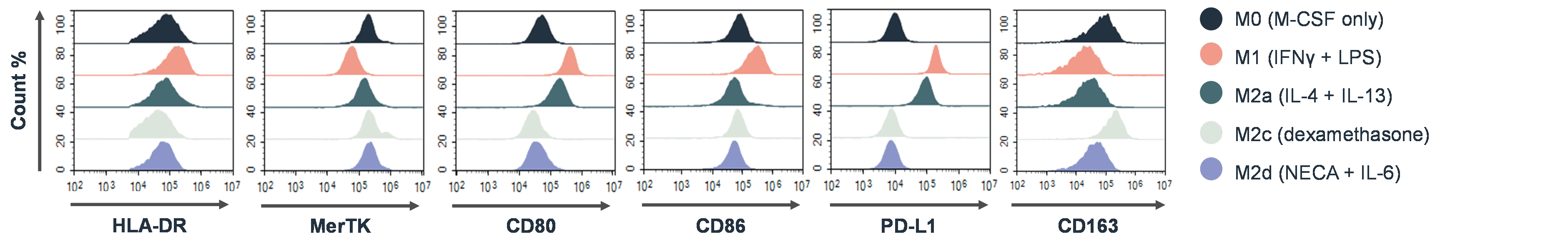
Macrophage polarisation autoimmune assays determine how therapeutics drive pro-regulatory macrophage states
In autoimmunity research, macrophage polarisation assays are used to study the functional states of macrophages, specifically their polarisation from a pro-inflammatory (M1) to an anti-inflammatory (M2) phenotype. These assays involve culturing macrophages with a therapeutic molecule under specific conditions to determine if M1 macrophages can be inhibited or redirected to an anti-inflammatory state. Phenotypic changes are assessed via altered cytokine production and changes in the expression of different surface markers. An example of flow cytometry data that would be used to determine macrophage polarisation states is shown below:
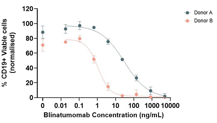
B cell depletion autoimmune assays quantify target B cell removal from PBMC or bone marrow matrices to support development of B cell–directed immunotherapies.
B cell depletion assays are used to evaluate the effectiveness of therapies that target B cells in cancers as well as autoimmune diseases. Current therapies used for this purpose include rituximab, which removes the autoantibody producing B cells in RA.
In B cell depletion assays PBMCs or bone marrow are incubated with therapeutic agents for a pre-determined time. The reduction in specific B cell populations is then measured via flow cytometry. An example of the type of data generated from a B cell depletion assay using the CD3xCD19 T cell engager blinatumomab is shown here:
T cell depletion autoimmune assays assess selective or broad T cell reduction using Fc-mediated effector mechanisms, including ADCC and ADCP, to characterise depletion strategies in autoimmune indications.
Specific rare autoreactive T cell populations represent attractive targets in T cell–driven autoimmune diseases. RoukenBio can assess T cell depletion using either pan T cells or defined rare T cell populations within Fc-mediated effector function assays (ADCC, ADCP) and TDCC.
RoukenBio, the Immunotherapy CRO redefined, delivers autoimmunity therapeutic development services that combine translational immunology expertise with human primary cell platforms and bespoke assay engineering, so you can generate decision-grade data with confidence.
Our advisory collaborative approach, backed by decades of collective experience of our brilliant minds, has led to the creation of an array of in vitro assay systems that recreate autoimmune environments, closely mirroring physiological processes.
We leverage our brilliant minds’ academic and industrial knowledge of autoimmunity to answer your specific questions.
Our assays are tailored to your specific needs.
Advanced flow cytometry, impedance-based systems, bioassays, and reporter-based technologies.
Let us help accelerate your autoimmunity research and therapeutic development with our bioassay expertise supporting your next breakthrough in autoimmunity treatment.


Autoimmune assays are in vitro tests that model loss of immune tolerance and allow you to study how immune cells behave in autoimmune settings. They measure functional immune responses such as T cell attenuation, cytokine secretion, depletion of specific B cell or T cell populations, and macrophage or regulatory T cell activity. They are used to quantify disease‑relevant inflammatory and regulatory pathways and how therapeutics modulate them.
Visual resources: Selected scientific illustrations on this website were created with BioRender.com and incorporate licensed BioRender content.