RoukenBio has established an in vitro T cell exhaustion model that can be used to assess candidate immunotherapeutics. Find out more about our unique approach.
Lynne Dunsford
Senior Study Manager

 Technical Presentations
|
October 20, 2022
|
5 min read
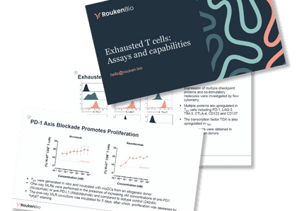
RoukenBio was the first CRO to develop an in vitro exhaustion process that yields a T cell phenotype that effectively mimics the look and behaviour of tumour infiltrating cells (TILs) found in the tumour microenvironment. Our in vitro exhausted T cells, generated from healthy human donor T cells, can be employed for the screening of checkpoint inhibitors, bispecific T cell engagers and multi-functional molecules and other immune modulators, including small molecules.
When exhausted T cells are functionally tested in a one-way mixed lymphocyte reaction (MLR) – their hypo-responsiveness is evident. Inclusion of checkpoint inhibitors in exhausted T cell, one-way MLRs demonstrates one of the key mechanisms of this class of molecules, which is the ability to overcome the chronic activation signals TILs are subject to in the tumour microenvironment and rejuvenate their function, inducing tumour immunity.
One of the major advantages of our in vitro model of human T cell exhaustion is the flexible assay set-up that allows multiple and customisable readouts, catered to specific scientific questions. A medium throughput set up, providing high content information in terms of phenotype (flow cytometry and cytokine production) and function; cytotoxicity, T cell activation, and cytokine secretion.
Inclusion of a benchmark molecule, the anti-PD-1 antibody Nivolumb, provides positive control for assay performance and gives confidence in the conclusions drawn from the data generated.
This results in a highly adaptable assay that is ideal for MoA assessment of checkpoint inhibitors, co- stimulation agonists and bispecific T cell engagers too. The combination of our knowhow and experience, coupled with exclusive in-house systems, technologies and donor pre-screening performed in advance, allows us to generate robust, quality data.
Initial studies demonstrating T cell exhaustion employed mouse models of chronic viral infections (e.g. LCMV). These are technically difficult and expensive to perform and are in a mouse background. Other traditional T cell activation assays employing polyclonal stimuli, e.g. SEB-activation, do not generate truly exhausted T cells and are therefore not as suitable to assess whether a compound has an impact on the reversal or inhibition of T cell exhaustion.
Therefore, it is recommended that you consider more applicable novel methods for the investigation of checkpoint inhibitors and other T cell function modulators.

Access the Capabilities brochure
🗓️ Stay informed with our monthly scientific newsletter, published on LinkedIn on the last Wednesday of each month.
These editions bring you the latest in drug development breakthroughs, industry trends, and expert insights from the brilliant minds at RoukenBio.
Subscribe today on LinkedInDelve into real data in our T cell exhaustion slide deck put together by our brilliant minds. Explore how our model recapitulates this exhausted state.

 Webinars
 Whitepapers