The advancement of immunotherapies, including CAR-T cell treatments, has driven a growing need for more predictive preclinical models. Addressing this, RoukenBio and ScreenIn3D have partnered to deliver next-generation 3D in vitro platforms that better replicate the tumour microenvironment.

Jamie Hodgson
VP of Business Development

 Research Posters
|
June 27, 2025
|
6 min read
Chimeric Antigen Receptor T-cell (CAR-T) therapies are approved for the treatment of several haematological malignancies. However, their clinical success against the majority of solid tumour types has been limited thus far. The need for physiologically relevant, more complex in vitro models of cancer is steadily increasing due to the emergence of immunotherapeutic approaches such as cell therapies but also drugs that actively target the immune system within the tumour microenvironment (TME). Furthermore, an increasing interest in precision treatment of cancer patients and the rise of combination of anti-cancer therapies has highlighted a gap for 3D tumour models in efficacy tests. Miniaturization facilitates increased data generation, especially when using clinical samples.
To meet this growing demand, RoukenBio, the Immunology CRO redefined, has formed a strategic partnership with ScreenIn3D, bringing together RoukenBio’s expertise in immunology and bespoke assay development with ScreenIn3D’s cutting-edge 3D miniaturized microfluidic technology. This collaboration enables us to deliver advanced, physiologically relevant assays that accelerate and de-risk your drug discovery efforts.
As immunotherapies and drugs targeting the tumour microenvironment (TME) gain momentum, there is a growing need for in vitro cancer models that more accurately reflect human pathology. Traditional 2D cell cultures often fail to capture the complexity of cell–cell and cell–matrix interactions within the TME, limiting their predictive value in drug discovery and development.
To address this, we are turning to advanced 3D in vitro models that better replicate the structural and functional characteristics of solid tumours. These physiologically relevant systems enable more accurate assessment of immune cell infiltration, drug penetration, and tumour-immune cell dynamics which ultimately enhance the translational value of preclinical studies and accelerate the development of next-generation cancer therapies.
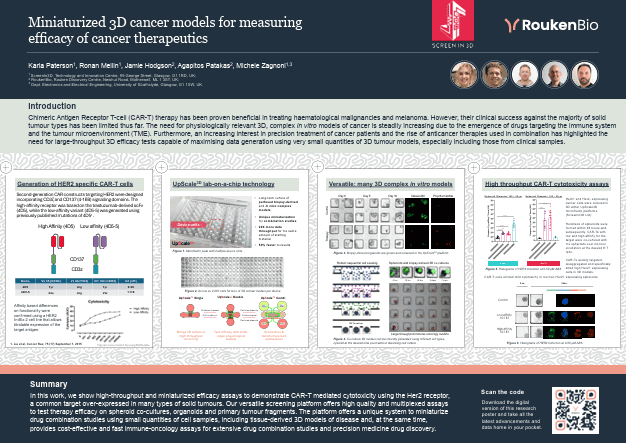
HER2-specific CAR-T cells are genetically engineered T cells that express a chimeric antigen receptor (CAR) designed to recognise and kill cancer cells that overexpress HER2 (human epidermal growth factor receptor 2). HER2 is commonly overexpressed in several solid tumours, including breast, ovarian, gastric, and lung cancers, making it a valuable therapeutic target.
Advantages of HER2 as a therapeutic target:
Challenges and Considerations:

Second-generation CAR constructs targeting HER2 were designed incorporating CD3ζ (signal 1) and CD137/4-1BB (signal 2) domains. The high-affinity receptor was based on the trastuzumab-derived scFv (4D5), while the low-affinity variant (4D5-5) was generated using previously published mutations of 4D51.

Affinity-based differences on functionality were confirmed using a HER2 inducible IndEx-2 cell line that allows titratable expression of the target antigen.
UpScale3D is a lab-on-a-chip technology developed by ScreenIn3D, our strategic partner. It focuses on miniaturizing and screening 3D disease models for drug discovery. The technology combines scalability with physiologically relevant microenvironments to assess molecule efficacy and improve candidate selection.

Figure 1 Microfluidic plate with multiple device units

Figure 2 As low as 2,000 cells for tens of 3D cellular models per device

High-throughput CAR-T cytotoxicity assays were used to measure how well CAR-T cells kill cancer cells.

Figure 5 Histograms of HER2 Induction at 0-50 μM ABA
HER2+ and HER2- expressing cancer cells were cultured in 3D within UpScale3D microfluidic platforms (ScreenIn3D).
Hundreds of spheroids were formed within 48 hours and, subsequently, CAR-Ts with low and high affinity for the target were co-cultured with the spheroids over 24-hour incubation at the desired E:T ratio.
CAR-Ts quickly targeted, disaggregated and specifically killed high HER2+ expressing cells in 3D models.
CAR-T cells elicited mild cytotoxicity in low/non-HER2+ expressing spheroids.

Figure 6 Histograms of HER2 Induction at 0-50 μM ABA
In summary, we showed how high-throughput and miniaturized efficacy assays can demonstrate CAR-T mediated cytotoxicity using the HER2 receptor, a common target over-expressed in many types of solid tumours. Our versatile screening platform offers high quality and multiplexed assays to test therapy efficacy on spheroid co-cultures, organoids and primary tumour fragments. The platform offers a unique system to miniaturize drug combination studies using small quantities of cell samples, including tissue-derived 3D models of disease and, at the same time, provides cost-effective and fast immune-oncology assays for extensive drug combination studies and precision medicine drug discovery.
References:
1 Liu et al, CancerRes; 75(17) September 1,2015.
Special thanks to Karla Paterson, Ronan Mellin and Michele Zagnoni from ScreenIn3D who helped in the creation of our collaborative research poster presented at AACR.
Figures were created by using BioRender.

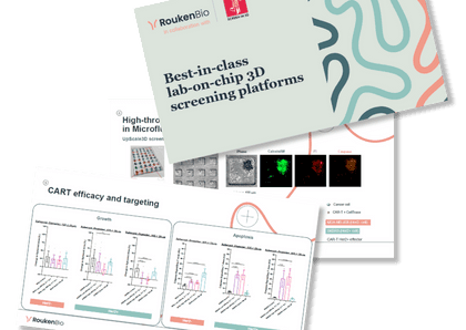
Access our Capabilities brochure
🗓️ Stay informed with our monthly scientific newsletter, published on LinkedIn on the last Wednesday of each month.
These editions bring you the latest in drug development breakthroughs, industry trends, and expert insights from the brilliant minds at RoukenBio.
Subscribe today on LinkedInIn partnership with Screenin3D, we deliver advanced 3D cell platforms that transform your research with cutting-edge, physiologically relevant miniaturized 3D cancer models.

 Insights
 Webinars