
Dr Luke Muir
Associate Scientific Director

Hannah Findlay
Senior Innovation Scientist
Dr Emilio Cosimo
Scientific Director of Innovation

 Research Posters
|
October 28, 2026
|
5 min read
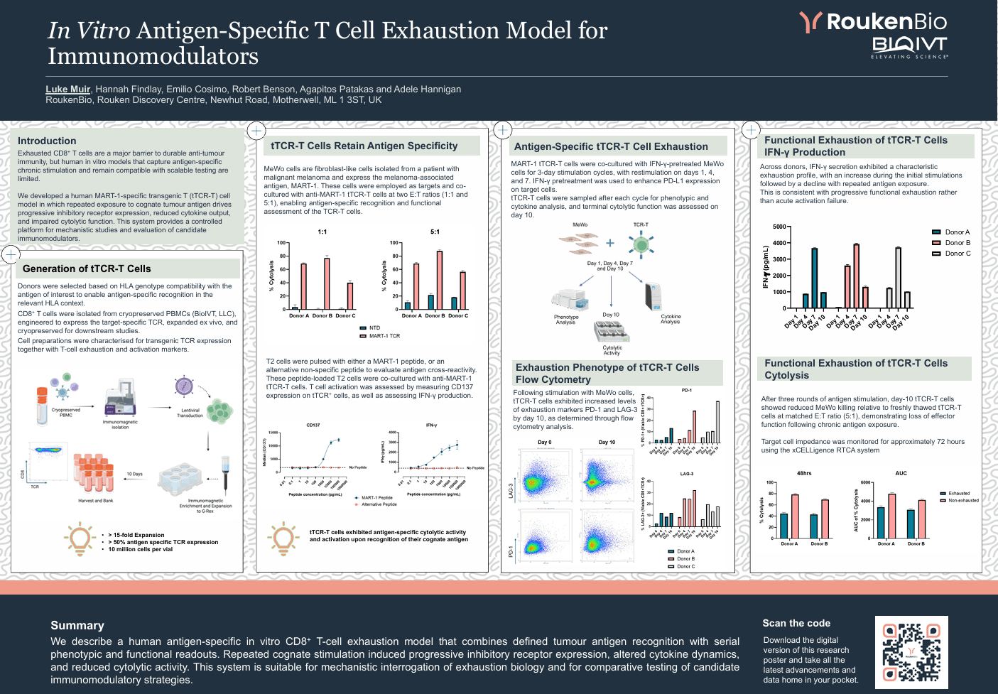
Exhausted CD8+ T cells are a major barrier to durable anti-tumour immunity, but human in vitro models that capture antigen-specific chronic stimulation and remain compatible with scalable testing are limited.
We developed a human MART-1-specific transgenic T (tTCR-T) cell model in which repeated exposure to cognate tumour antigen drives progressive inhibitory receptor expression, reduced cytokine output, and impaired cytolytic function. This system provides a controlled platform for mechanistic studies and evaluation of candidate immunomodulators.
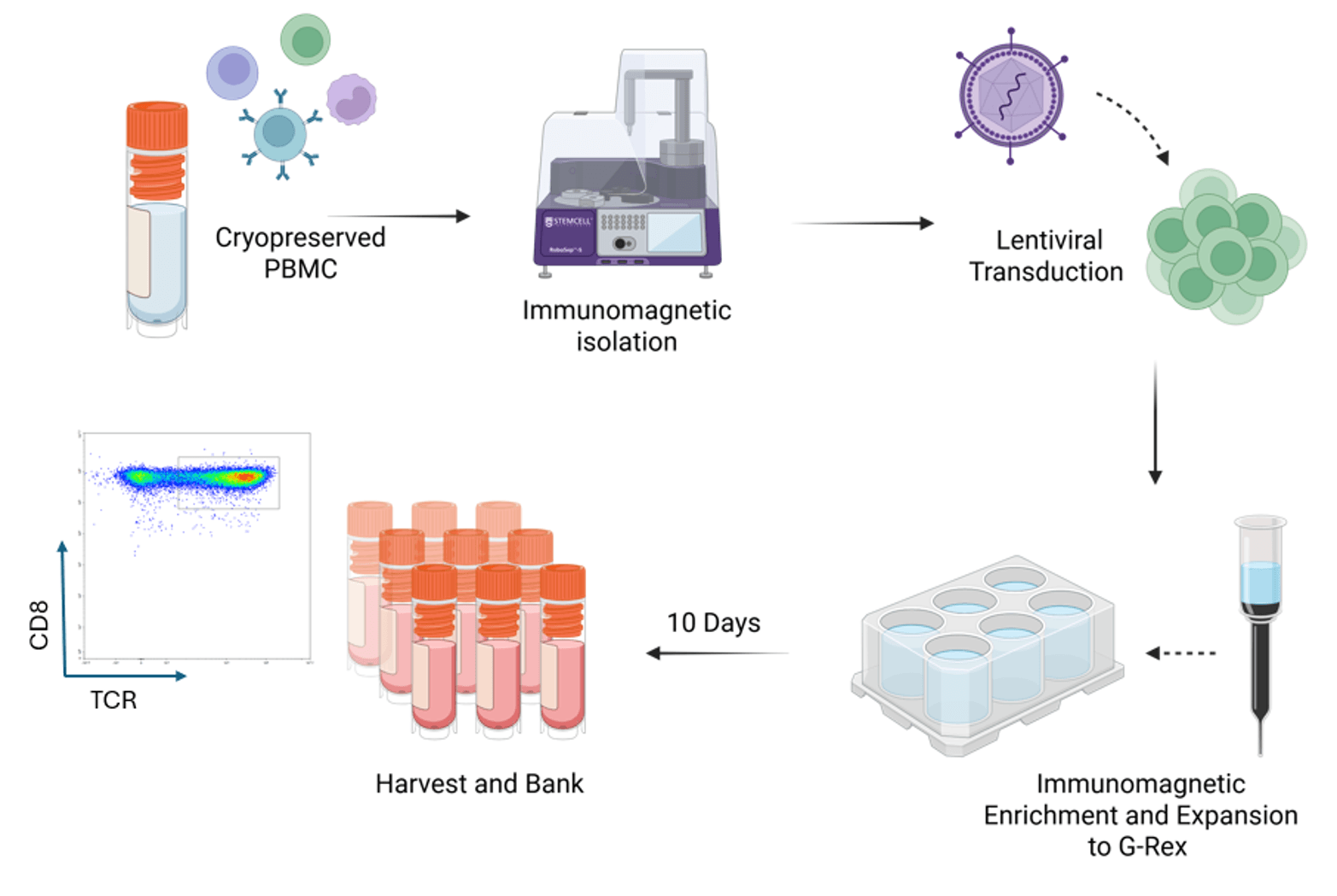
Donors were selected based on HLA genotype compatibility with the antigen of interest to enable antigen-specific recognition in the relevant HLA context.
CD8+ T cells were isolated from cryopreserved PBMCs (BioIVT, LLC), engineered to express the target-specific TCR, expanded ex vivo, and cryopreserved for downstream studies.
Cell preparations were characterised for transgenic TCR expression together with T-cell exhaustion and activation markers.
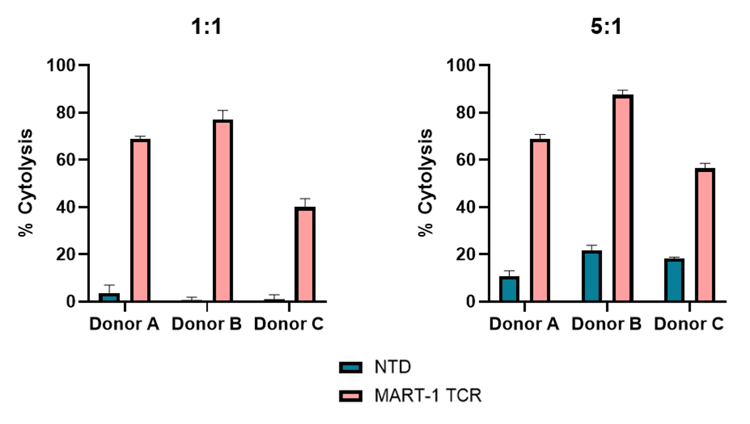
MeWo cells are fibroblast-like cells isolated from a patient with malignant melanoma and express the melanoma-associated antigen, MART-1. These cells were employed as targets and co-cultured with anti-MART-1 tTCR-T cells at two E:T ratios (1:1 and 5:1), enabling antigen-specific recognition and functional assessment of the TCR-T cells.
T2 cells were pulsed with either a MART-1 peptide, or an alternative non-specific peptide to evaluate antigen cross-reactivity. These peptide-loaded T2 cells were co-cultured with anti-MART-1 tTCR-T cells. T cell activation was assessed by measuring CD137 expression on tTCR+ cells, as well as assessing IFN-γ production
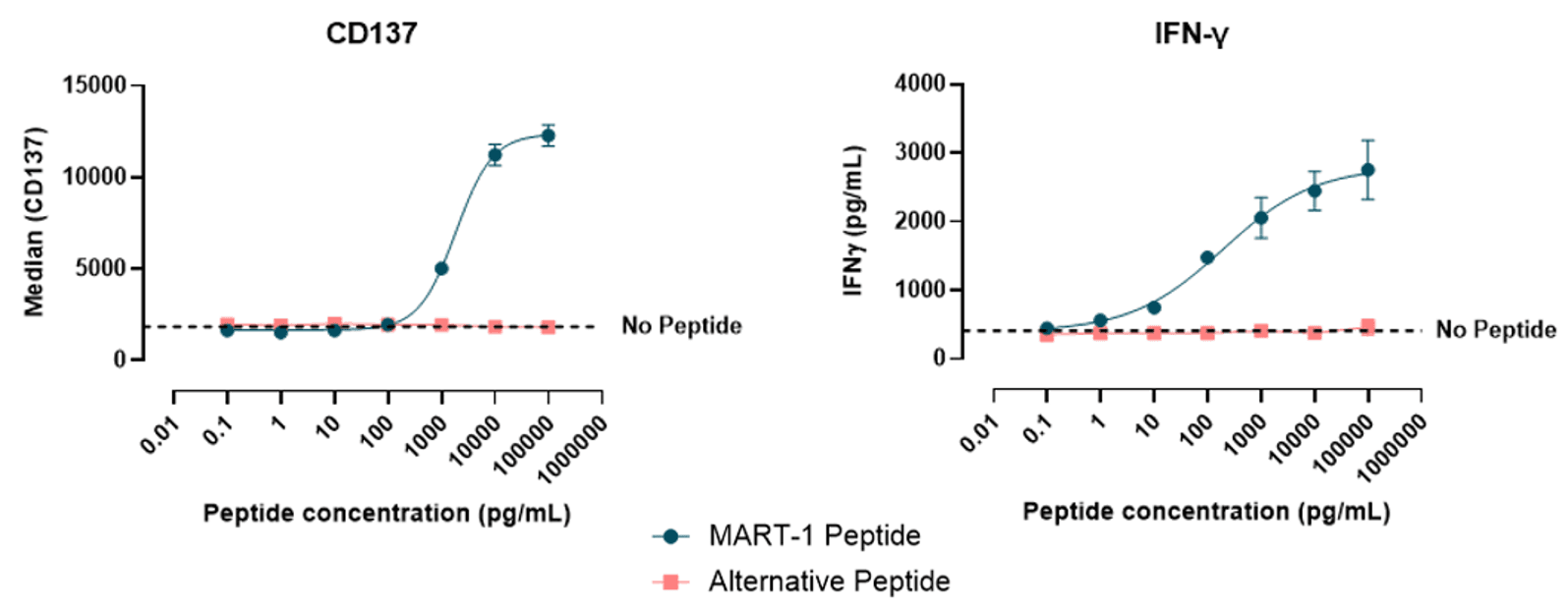
tTCR‑T cells exhibited antigen‑specific cytolytic activity and activation upon recognition of their cognate antigen
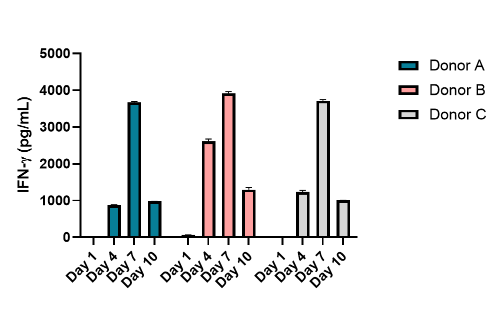
MART-1 tTCR-T cells were co-cultured with IFN-γ-pretreated MeWo cells for 3-day stimulation cycles, with restimulation on days 1, 4, and 7. IFN-γ pretreatment was used to enhance PD-L1 expression on target cells.
tTCR-T cells were sampled after each cycle for phenotypic and cytokine analysis, and terminal cytolytic function was assessed on day 10.
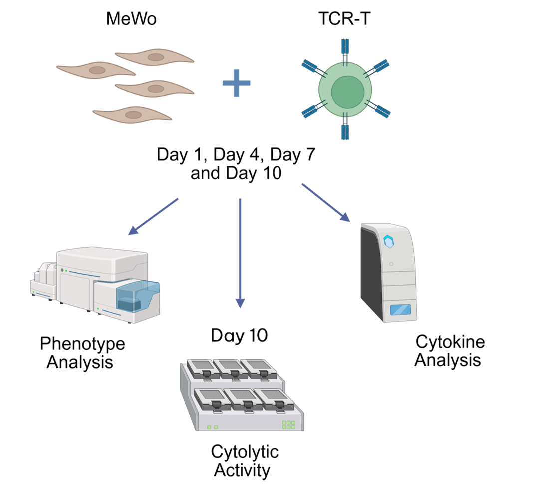
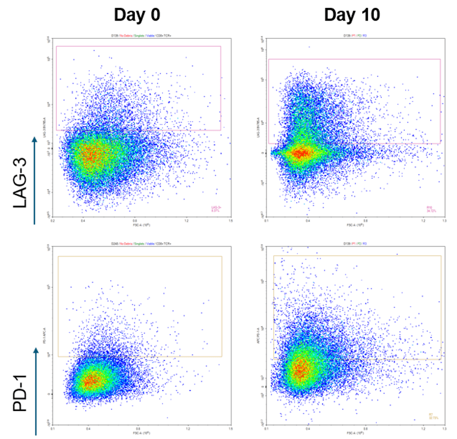
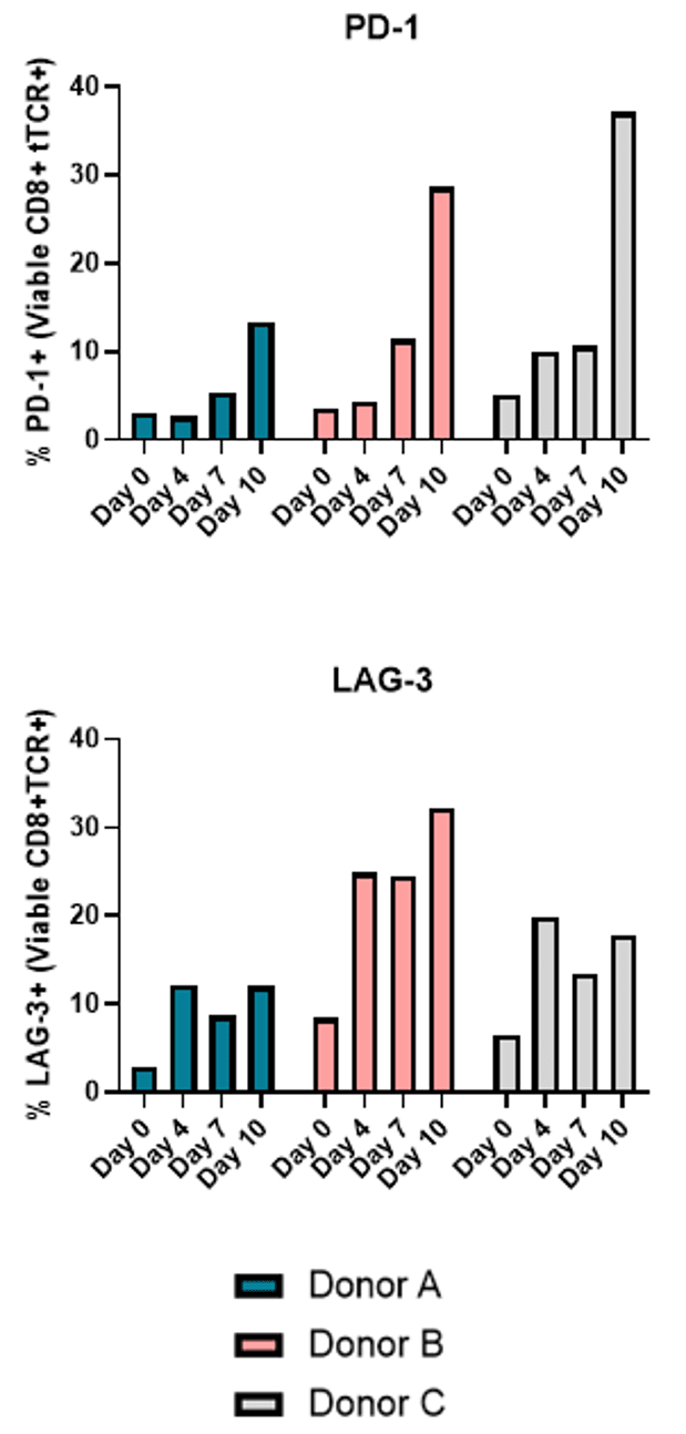
Following stimulation with MeWo cells, tTCR‑T cells exhibited increased levels of exhaustion markers PD‑1 and LAG‑3 by day 10, as determined through flow cytometry analysis.
Across donors, IFN-γ secretion exhibited a characteristic exhaustion profile, with an increase during the initial stimulations followed by a decline with repeated antigen exposure.
This is consistent with progressive functional exhaustion rather than acute activation failure.
After three rounds of antigen stimulation, day-10 tTCR-T cells showed reduced MeWo killing relative to freshly thawed tTCR-T cells at matched E:T ratio (5:1), demonstrating loss of effector function following chronic antigen exposure.
Target cell impedance was monitored for approximately 72 hours using the xCELLigence RTCA system
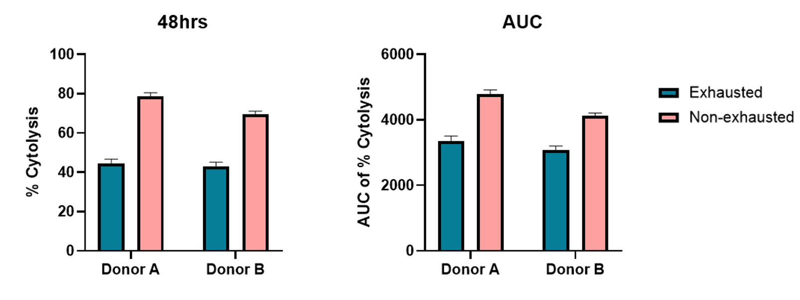
We describe a human antigen-specific in vitro CD8+ T-cell exhaustion model that combines defined tumour antigen recognition with serial phenotypic and functional readouts. Repeated cognate stimulation induced progressive inhibitory receptor expression, altered cytokine dynamics, and reduced cytolytic activity. This system is suitable for mechanistic interrogation of exhaustion biology and for comparative testing of candidate immunomodulatory strategies.
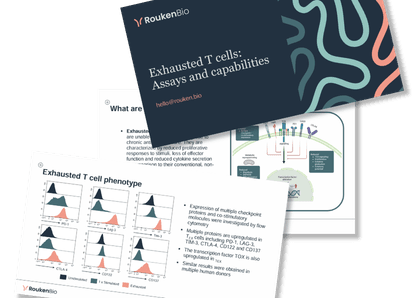
Access the slides
🗓️ Stay informed with our monthly scientific newsletter, published on LinkedIn on the last Wednesday of each month.
These editions bring you the latest in drug development breakthroughs, industry trends, and expert insights from the brilliant minds at RoukenBio.
Subscribe today on LinkedInDiscover our human in vitro T-cell exhaustion model and explore how it enables mechanistic studies and evaluation of candidate immunomodulators.


 Insights
 Webinars
 Technical Presentations