Dendritic Cell Assays
We offer a range of dendritic cell (DC) assays to support your immunological research and therapeutic development, using monocyte-derived DCs or where appropriate DCs isolated from PBMCs. Each dendritic cell assay provides comprehensive insights into DC functionality, activation, and their role in immune responses.

Driving the immune response
Dendritic cells (DCs) are specialised cells of the immune system bridging the gap between innate and adaptive immunity. As professional antigen presenting cells (APCs), DCs are responsible for initiating and driving immune responses to pathogens and cancer cells whilst maintaining tolerance to environmental and self-antigens. The balance between immunogenic and tolerogenic states can be disrupted in various diseases where immunogenic DCs drive autoimmunity whilst tolerogenic DCs allow tumour growth and metastasis in cancer.

Explore our Dendritic Cell Assays
Our dendritic cell assay formats help researchers investigate this balance and understand DC behaviour in diverse immune contexts.
DC differentiation
DC Activation
Mixed Lymphocyte Reaction (MLR)
Antigen Presentation to T Cells
DC differentiation
At RoukenBio, we have developed a standardised protocol for the generation of DCs. CD14+ monocytes are magnetically isolated from cryopreserved PBMCs and incubated with GM-CSF and IL-4 over a 7-day period. The phenotype and purity of these moDCs is confirmed by flow cytometry after which the cells can be used in assays. Where appropriate magnetic isolation techniques can be applied to isolate DCs from PBMCs directly however this is limited due to the low recovery rate.
This workflow supports any dendritic cell assay requiring controlled, reproducible DC differentiation.
DC Activation
DCs become activated when exposed to certain soluble mediators (such as TLR agonists) in the inflammatory environment. This leads to an upregulation of MHCII, costimulatory molecules (CD80/86) and the release of inflammatory cytokines. In DC activation assays, DCs are incubated with a stimulus in the presence of a candidate therapeutic with flow cytometry and cytokine analysis used to assess activation.
These dendritic cell assay formats are well suited for screening candidate therapeutics for their ability to drive or repress DC activation.
Mixed Lymphocyte Reaction (MLR)
The ability of DCs to activate T cells can be assessed using a mixed lymphocyte reaction (MLR). In this assay, naïve or mature DCs are incubated with T cells from an allogenic donor. The HLA mismatch between donors drives T cell activation, which can be assessed by flow cytometry and cytokine analysis. Candidate therapeutics that alter the activation state of DCs can be readily tested within this system, making this a valuable dendritic cell assay for immunomodulatory screening.
Human donor selection
Donor pairing is crucial for effective MLR. With an onsite bank of ~70 recallable cryopreserved PBMCs and three onsite phlebotomists, RoukenBio can regularly screen donor pairs to ensure optimal assay conditions. Pairings in which Nivolumab can enhance the levels of INFγ production in a dose-dependent manner provide favourable pairings for assessment of test articles.

Pre-screening of donor combinations - four PBMC donors (A-D), 12 allogenic combinations, INFγ ELISA. Donor pairs driving a dose-dependent increase in INFγ production are selected for our Customer studies.
Mixed lymphocyte reaction readouts:

Additional readouts can be included in an MLR based upon the needs of your study including T cell proliferation, assessment of activation markers and the measurement of additional cytokines aside from INFγ.
Antigen Presentation to T Cells
Antigen presentation can be indirectly assessed using common pathogen derived (CMV, tetanus toxoid, Candida albicans) or cancer (MART-1) antigens. DCs are generated from appropriate donors and are pulsed with the relevant antigen in the presence of a candidate therapeutic prior to co-culture with T cells from the same donor. T cell activation, as measured by flow cytometry and cytokine analysis, acts an indirect indicator of antigen presentation. Alternatively, reporter cell lines, such as NFAT reporters can be used with luminescence acting as an indication of T cell activation and therefore antigen presentation.
Candidate therapeutics that influence antigen presentation can be readily assessed within this system. This provides a robust dendritic cell assay setup for evaluating antigen-specific responses.
Discover our in vitro MLR models
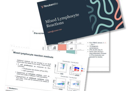
Our one-way and two-way MLR assays use pre-screened human T cells co-cultured with antigen presenting cells (APCs). Explore more details in our presentation.
Access our MLR technical slide deck
