RoukenBio is a specialist immunology CRO with extensive experience in developing mechanism-driven, translational in vitro models designed to characterise target engagement, potency, mechanism of action and functional activity across allergic and type 2 inflammatory pathways.
From established pathways to novel targets, we design fit-for-purpose assays around your therapeutic modality and biological question.

RoukenBio is a specialist immunology CRO providing human-relevant in vitro assay development and testing to support the discovery and preclinical development of allergy and type 2 inflammation therapeutics. We design mechanism-driven, translational assay strategies that generate robust, decision-enabling data across biologics, small molecules, cell therapies and emerging therapeutic modalities.
Our platform encompasses the interconnected epithelial, innate and adaptive immune pathways that drive allergic and type 2 inflammation. This includes IgE/FcεRI signalling, mast cell and basophil activation, eosinophil function, Th2 cytokine biology, B cell responses and immune cell crosstalk, alongside upstream epithelial alarmin pathways including TSLP, IL-33 and IL-25.
We develop and optimise assays using primary human immune cells, engineered reporter systems, co-culture models and differentiated tissue-relevant systems, selecting the most appropriate model according to the therapeutic target, modality and biological question. Our platforms incorporate key cellular drivers of allergic inflammation, including mast cells, basophils, eosinophils, T cells, Th2 cells, regulatory T cells and B cells, enabling investigation of pathway specific and integrated immune responses.
These human in vitro models support assessment of target engagement, cellular potency, mechanism of action, IgE-mediated activation, cytokine and chemokine responses, degranulation, immune cell function and therapeutic inhibition. By progressing from defined pathway assays through primary cell and more complex tissue relevant models, we help generate an integrated evidence package to support candidate selection, mechanistic understanding and translational development.

Allergic and type 2 inflammatory diseases result from complex interactions between epithelial, innate and adaptive immune pathways. RoukenBio translates this biology into fit-for-purpose cellular and tissue models spanning the key stages of the inflammatory cascade from epithelial alarmin release and type 2 cytokine signalling through IgE/FcεRI-mediated activation and downstream effector cell responses.
Our capabilities support programmes targeting established and emerging pathways, including IgE/FcεRI, IL-4, IL-5, IL-13, TSLP, IL-33 and IL-25, as well as downstream mast cell, basophil, eosinophil and other immune cell functions.
From target engagement and potency to mechanism of action and functional efficacy, RoukenBio provides integrated, human-relevant assay strategies to help advance the next generation of allergy and type 2 inflammation therapeutics.

RoukenBio combines specialist immunology expertise with human relevant cellular and tissue models to support therapeutic development across allergic and type 2 inflammatory diseases. Our integrated approach enables investigation of disease relevant pathways from receptor signalling and immune cell activation through to downstream functional and tissue responses.
Asthma and Airway Inflammation
Food and IgE Mediated Allergy
Atopic and Type 2 Inflammatory Disease
Emerging Targets and Mechanisms
Explore mechanisms driving type 2 airway inflammation using complementary immune cell and differentiated airway epithelial models. Our platforms enable investigation of TSLP, IL 4 and IL 13 signalling, inflammatory biomarker responses, mucus production and epithelial remodelling to support the development of therapies targeting asthma and related airway disease.
Explore mechanisms driving type 2 airway inflammation using complementary immune cell and differentiated airway epithelial models. Our platforms enable investigation of TSLP, IL 4 and IL 13 signalling, inflammatory biomarker responses, mucus production and epithelial remodelling to support the development of therapies targeting asthma and related airway disease.
Characterise allergen driven and IgE dependent responses using models spanning IgE and FcεRI signalling, basophil activation, mast cell degranulation and allergen specific immune responses. These complementary approaches support assessment of therapeutic potency, mechanism of action and functional inhibition of allergic responses.
Investigate key pathways underlying type 2 inflammation using primary human immune cells, engineered reporter systems and functional assays. Models targeting IL 4, IL 13, TSLP and downstream biology provide translational approaches for evaluating therapeutic modulation of immune signalling and cellular function.
Our scientific team develops bespoke assay strategies for novel targets and therapeutic mechanisms in allergy and type 2 inflammation. By combining engineered reporter systems with primary human cells and tissue relevant models, we can build fit-for-purpose assay cascades that support target validation, candidate characterisation and mechanism of action studies.
Supporting biologics, bispecifics, small molecules and emerging therapeutic modalities from early target validation through to mechanistic and translational assessment.
Our assay strategies use the most appropriate cellular model for the biological question, therapeutic modality and development stage.
Th2 cells
B cells
Eosinophils
Mast cells
Basophils
Airway Epithelial Models
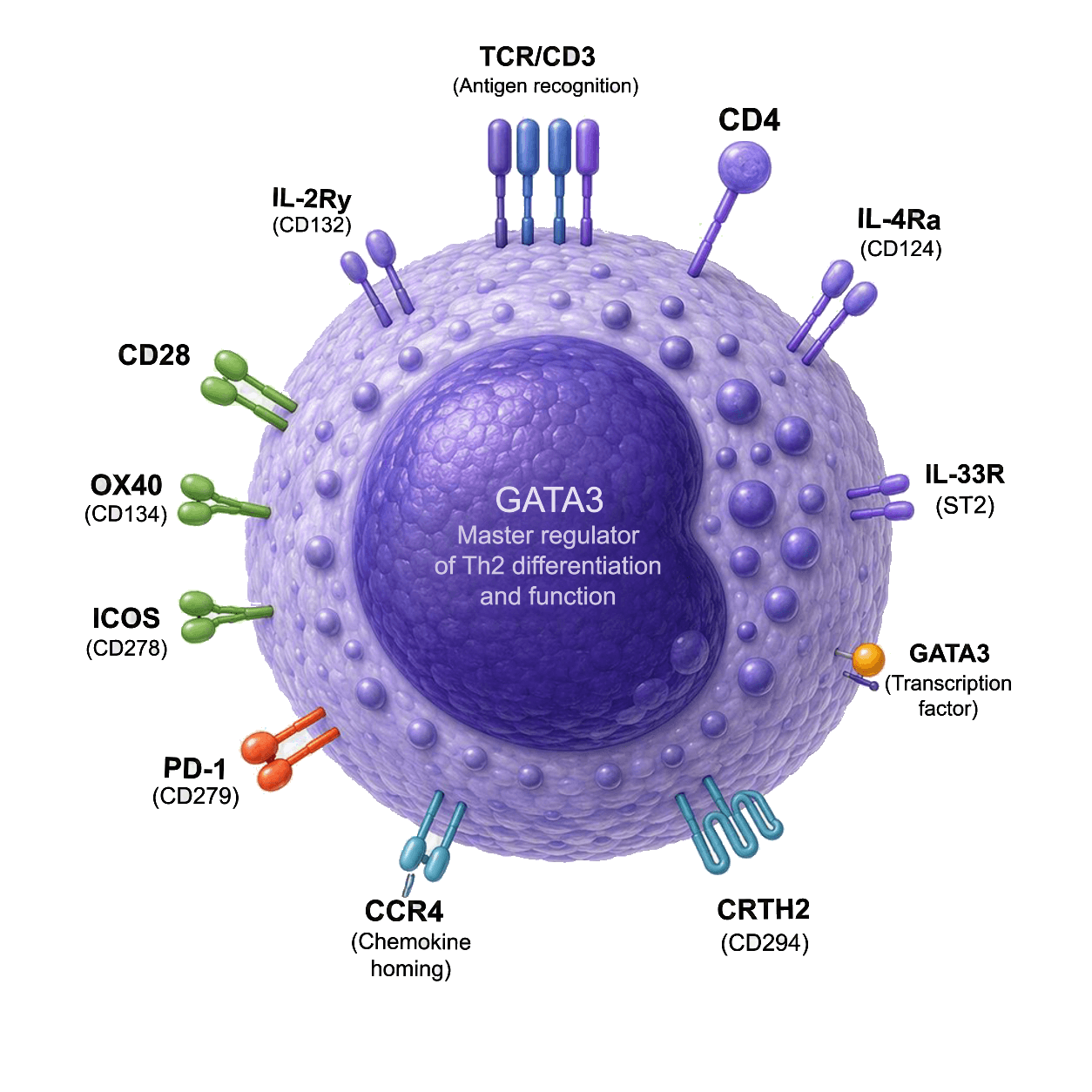
Th2 biology is central to type 2 inflammation and the induction of IgE class switching in B cells. Accordingly, Th2-relevant functional readouts are widely applied in allergy drug discovery to quantify cytokine modulation and to characterise downstream immunological effects on effector cell activation, antibody production, and inflammatory signalling pathways.
CD4 T helper (Th cells) choreographs adaptive immune responses. Th2 cells play a critical role in protective immunity against helminth infections; however, dysregulated Th2 responses also drive the pathogenic type 2 inflammation that characterises allergic disease. Their activation and secretion of typical Th2 cytokines (IL-4, IL-5, IL-9 and IL-13) drive B cell proliferation and immunoglobulin class-switching to IgE, eosinophilia and mastocytosis, goblet cell hyperplasia, alternative macrophage activation (M2 polarisation) and smooth muscle contraction, collectively contributing to type 2 immune responses.
Th2 biology is central to type 2 inflammation and the induction of IgE class switching in B cells. Accordingly, Th2-relevant functional readouts are widely applied in allergy drug discovery to quantify cytokine modulation and to characterise downstream immunological effects on effector cell activation, antibody production, and inflammatory signalling pathways.
CD4 T helper (Th cells) choreographs adaptive immune responses. Th2 cells play a critical role in protective immunity against helminth infections; however, dysregulated Th2 responses also drive the pathogenic type 2 inflammation that characterises allergic disease. Their activation and secretion of typical Th2 cytokines (IL-4, IL-5, IL-9 and IL-13) drive B cell proliferation and immunoglobulin class-switching to IgE, eosinophilia and mastocytosis, goblet cell hyperplasia, alternative macrophage activation (M2 polarisation) and smooth muscle contraction, collectively contributing to type 2 immune responses.

B cells play a central role in allergic sensitisation through the production of allergen-specific IgE. Th2-derived signals, particularly IL-4 and IL-13, drive B cell activation and immunoglobulin class switching towards IgE. Allergen specific IgE subsequently sensitises mast cells and basophils through FcεRI, linking adaptive immune responses to downstream allergic effector cell activation. B cell assays can therefore provide mechanistic insight into IgE production, class switching and therapeutic modulation of the allergic response.
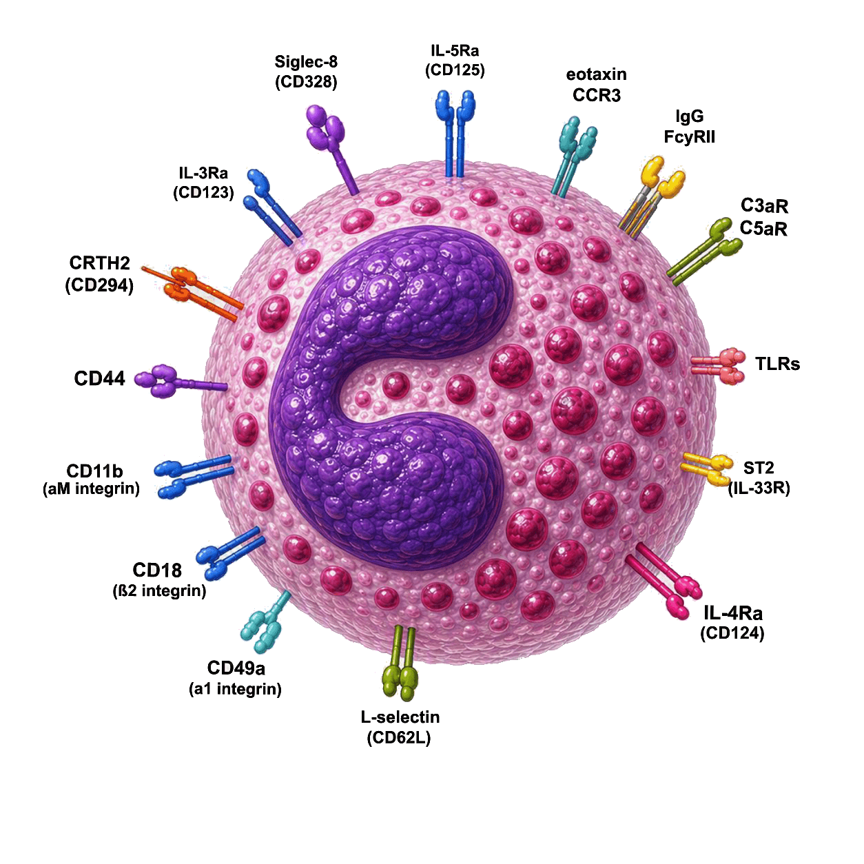
Eosinophils are key drivers of tissue inflammation in allergic disease, making them highly relevant for allergy assay design and profiling of immune-modulating therapeutics.
Upon activation, eosinophils release a host of cytotoxic proteins including major basic protein (MBP), eosinophil cationic protein (ECP), eosinophil-derived neurotoxin (EDN) and eosinophil peroxidase (EPO). In addition, eosinophils can release a host of immunomodulatory cytokines and chemokines to shape adaptive immune responses.

Mast cells are central to many allergy assays because IgE crosslinking at FcεRI triggers rapid degranulation and mediator release (e.g., histamine, proteases, lipid mediators and cytokines). For allergy drug discovery, mast cell models enable direct evaluation of therapies designed to inhibit FcεRI signalling, mediator release and activation pathways.
A central event in acute allergic reactions is the triggering of mast cells, granulated-tissue-resident innate cells found in peripheral tissues such as skin, airways and gastrointestinal tract.
Antigen-specific activation of these mast cells occurs when surface bound IgE is crosslinked by binding of an allergen, driving activation of the mast cell via the high affinity IgE receptor (FεRI). Triggering in this way results in mast cell release of a wide range of proinflammatory and vasoactive mediators derived from preformed granules, arachidonic acid metabolites and as cytokines.
Key examples include histamine, proteases (e.g., tryptase, chymase) and enzymes (e.g., betahexosaminidase); prostaglandins and leukotrienes (e.g., PGD2, LTC4 respectively); and cytokines (e.g., IL-1β, IL-6). This ability to release a wide range of mediators, makes them key players in modulating the immune response and therefore mast cells and their products are attractive therapeutic targets.
A significant barrier to studying human mast cell activity arises from their tissue resident nature, making acquiring sufficient cell numbers for assay use challenging. RoukenBio offers in vitro mast cell differentiation as a valuable tool for use in functional assays for the screening of candidate therapeutics, as well as custom cell lines to monitor triggering of FεRI.
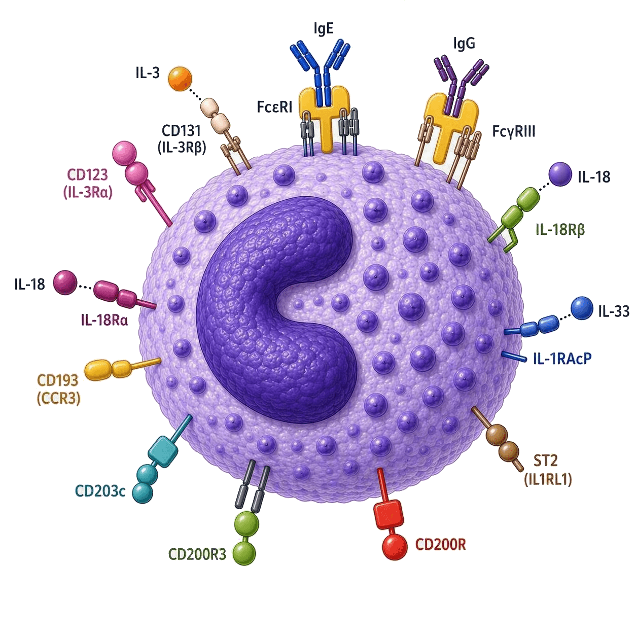
Basophils are circulating effector cells that contribute to the initiation and amplification of allergic inflammation. Through their high-affinity IgE receptor, FcεRI, basophils respond to allergen-mediated IgE crosslinking, releasing histamine and producing type 2 cytokines including IL-4 and IL-13. Their ability to rapidly amplify IgE-mediated and type 2 responses makes them an important target for allergy therapeutics and a valuable human primary-cell model for assessing basophil activation and therapeutic inhibition.
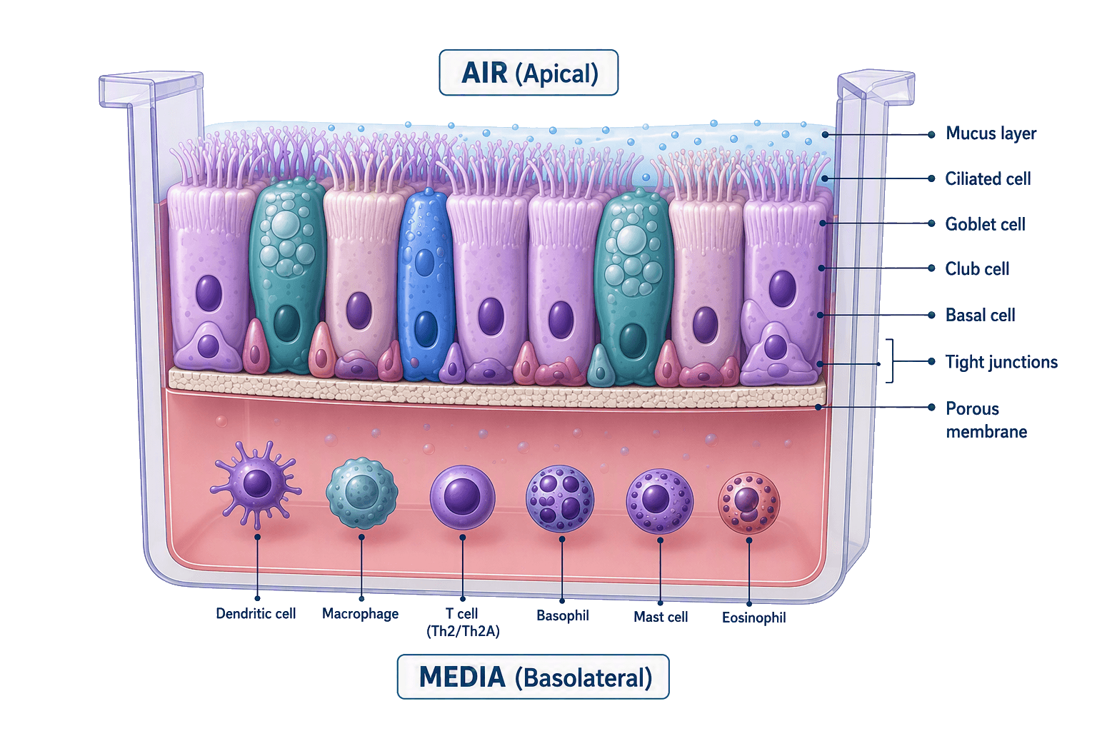
Differentiated human airway epithelial models provide a physiologically relevant system for investigating tissue responses associated with asthma and type 2 inflammation.
RoukenBio uses primary human bronchial epithelial cells cultured at the air liquid interface to recreate key features of the airway epithelium. Exposure to type 2 cytokines, including IL 4 and IL 13, enables assessment of disease relevant epithelial responses such as mucus production, epithelial remodelling and secretion of inflammatory biomarkers including CCL26.
These models complement primary immune cell and reporter assays by extending therapeutic assessment from pathway modulation to functional changes within a relevant human tissue system. Integration of imaging, histology and soluble biomarker endpoints enables evaluation of therapeutic activity and mechanism of action within a single experimental platform.
RoukenBio’s allergy assay platform combines primary human immune-cell models, engineered reporter systems and physiologically relevant airway models to interrogate allergic and type 2 inflammation across multiple levels of biology. From primary immune cell activation and allergen specific responses, through Th2 polarisation and IL-4/IL-13-driven cytokine biology, to downstream IgE/FcεRI-mediated effector responses such as mast cell and basophil degranulation, our assays enable therapeutic activity to be characterised across the allergic inflammatory cascade.
Differentiated air–liquid interface (ALI) airway epithelial models extend this approach to tissue relevant biology, enabling assessment of type 2 cytokine driven epithelial responses, including mucus production, airway remodelling and inflammatory biomarker release. Together, these complementary platforms provide readouts of pathway activity, target engagement and functional response to support mechanism-of-action studies, compound profiling and therapeutic development.
We can identify the appropriate assay or develop a bespoke model to address the critical biological questions for your therapeutic programme.
Assay/ Tool | What it measures | Application |
Primary immune cell activation | pSTAT6, CD23, TARC/CCL17 | Assessment of IL-4/IL-13 pathway inhibition and downstream biology |
Th2 cell assays | type 2 responses | Characterisation of drug effects on Th2 polarisation/ cytokine secretion |
Allergen recall assays | Antigen-specific T-cell/type 2 responses | Characterisation of drug effects on allergen driven adaptive immunity |
Degranulation assays | Histamine, β-hexosaminidase, tryptase and/or CD63, CD107 | Assessment of allergic effector cell activation and inhibition |
Basophil activation test (BAT) | CD63, CD203c | Evaluation of IgE-mediated activity in human blood |
Reporter cell lines | TSLP, IL-4/IL-13, FcεRI signalling | Rapid pathway profiling, mechanism of action and potency assessment |
Human Air liquid interface (ALI) airway model | Epithelium remodelling, mucus production, cytokine secretion | Evaluation of IL-4/IL-13 pathway inhibitors in airway epithelial models using functional and biomarker endpoints to support compound prioritisation. |
Primary Cell Assays | Peripheral Blood Mononuclear Cells
Primary Immune Cell Assays | B cells
Th2 Cell Assays
Allergen recall assays
Mast Cell Degranulation Assays
Basophil Activation Test (BAT)
Reporter cell lines
Human Air–Liquid Interface (ALI) Airway Model
Peripheral blood mononuclear cells (PBMCs) provide a physiologically relevant approach to understanding how therapeutic candidates modulate allergic and type 2 inflammatory pathways. Incorporating multiple immune cell populations, PBMC assays enable therapeutic activity to be assessed within a complex cellular environment while retaining clinically relevant donor biology. RoukenBio combines multiparameter flow cytometry and functional cytokine/chemokine readouts to capture both proximal signalling and downstream cellular responses.
RoukenBio’s PBMC assays enable interrogation of key type 2 inflammatory pathways using complementary proximal and downstream functional readouts. IL-4- and IL-13-driven responses can be characterised through STAT6 phosphorylation (pSTAT6), CD23 upregulation and TARC/CCL17 production, providing measures of pathway activation across different levels of the signalling cascade. These assays can be tailored to assess target engagement, pathway inhibition, compound potency and mechanism of action, while enabling evaluation of donor-to-donor variability in primary human cells.

Peripheral blood mononuclear cells (PBMCs) provide a physiologically relevant approach to understanding how therapeutic candidates modulate allergic and type 2 inflammatory pathways. Incorporating multiple immune cell populations, PBMC assays enable therapeutic activity to be assessed within a complex cellular environment while retaining clinically relevant donor biology. RoukenBio combines multiparameter flow cytometry and functional cytokine/chemokine readouts to capture both proximal signalling and downstream cellular responses.
RoukenBio’s PBMC assays enable interrogation of key type 2 inflammatory pathways using complementary proximal and downstream functional readouts. IL-4- and IL-13-driven responses can be characterised through STAT6 phosphorylation (pSTAT6), CD23 upregulation and TARC/CCL17 production, providing measures of pathway activation across different levels of the signalling cascade. These assays can be tailored to assess target engagement, pathway inhibition, compound potency and mechanism of action, while enabling evaluation of donor-to-donor variability in primary human cells.
B cells play a central role in allergic sensitisation through the generation of allergen-specific IgE and provide a critical link between adaptive immunity and downstream allergic effector responses. Following activation, type 2 cytokine signals, particularly IL-4, promote B cell differentiation and immunoglobulin class switching towards IgE. Secreted IgE subsequently sensitises mast cells and basophils through FcεRI, enabling rapid effector cell activation upon allergen re-exposure.
RoukenBio’s primary human B-cell assays enable functional interrogation of B cell activation, differentiation and IgE responses. Using PBMCs, isolated B cells or defined B-cell subsets, stimulation conditions can be tailored to investigate B cell activation, proliferation, antibody-secreting cell differentiation and IgE production. Multiparametric flow cytometry and soluble antibody measurements provide complementary cellular and functional readouts, enabling assessment of therapeutic modulation across different stages of the B cell response.
These assays support mechanism-of-action studies and compound profiling for therapeutics targeting B cell activation, type 2 cytokine signalling, IgE class switching or antibody production, providing a human-relevant platform for investigating the adaptive immune mechanisms underlying allergic disease.

Th2 cells are central drivers of allergic and type 2 inflammation, coordinating immune responses through the production of cytokines including IL-4, IL-5 and IL-13. These cytokines promote IgE class switching in B cells, eosinophil recruitment and activation, and tissue responses that contribute to the initiation and persistence of allergic disease.
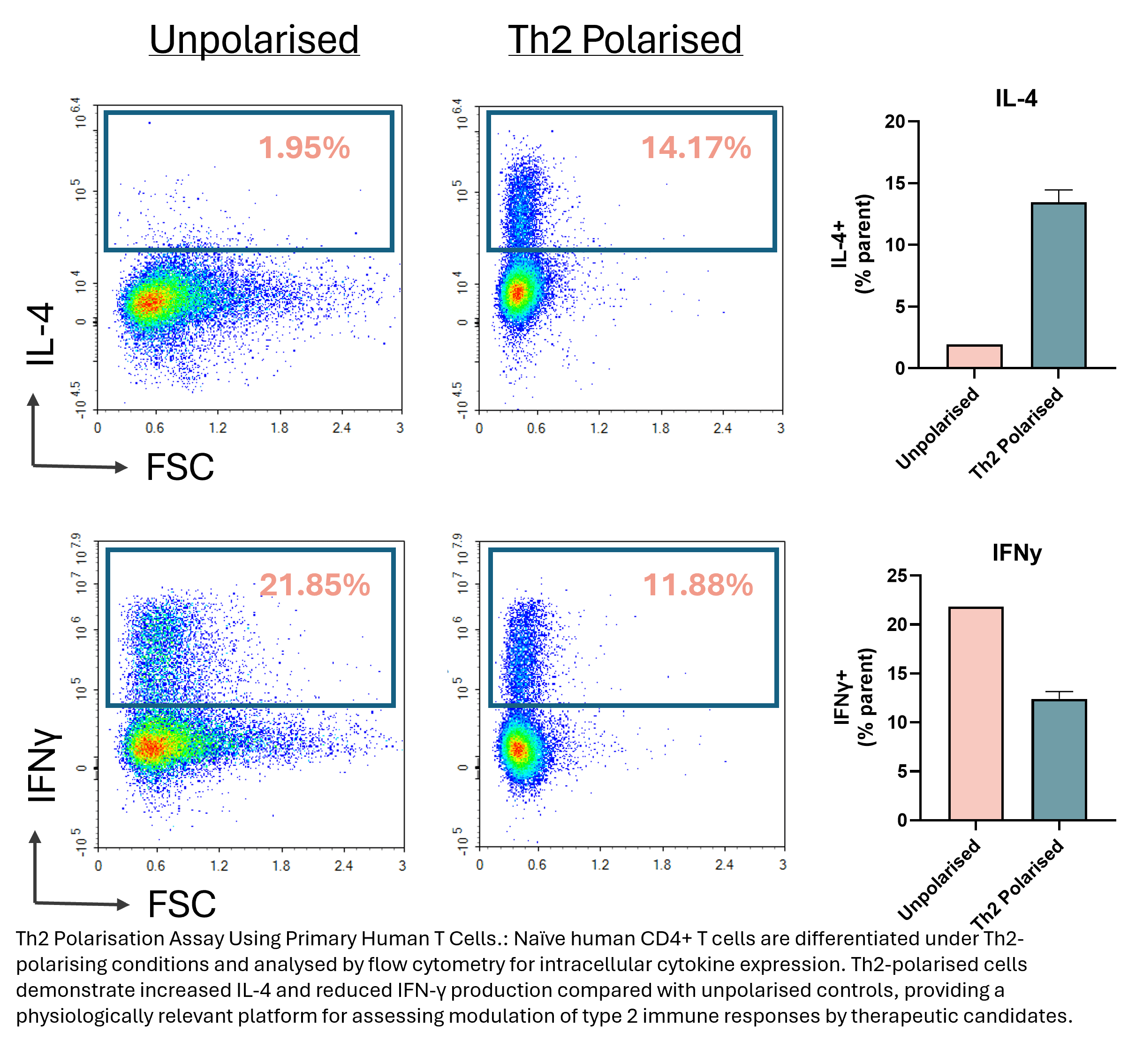
RoukenBio’s primary human Th2 cell assays enable functional interrogation of therapeutic modulation of Th2 differentiation and effector responses. Naïve CD4⁺ T cells can be differentiated under Th2 polarising conditions to assess the impact of candidate therapeutics on Th2 polarisation, proliferation and cytokine production. Multiparametric readouts, including phenotypic markers and secretion of IL-4, IL-5 and IL-13, enable characterisation of compound activity across different stages of the Th2 response. These assays support mechanism-of-action studies, compound profiling and evaluation of therapeutics targeting T cell activation, differentiation or downstream type 2 cytokine pathways, providing a physiologically relevant complement to engineered reporter systems.
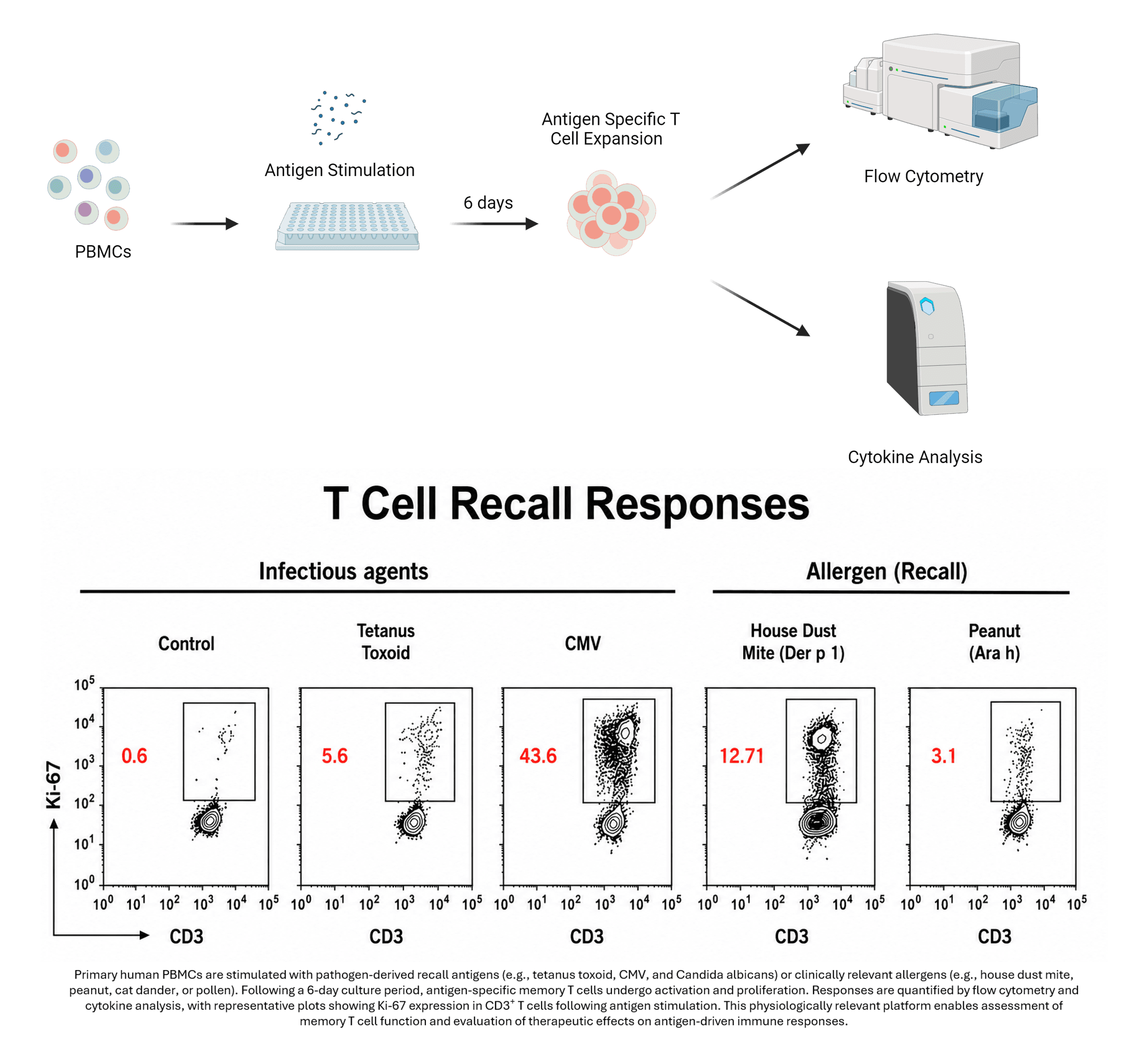
Allergen recall assays model the antigen-specific adaptive immune response that underpins allergic inflammation. Following previous sensitisation, allergen specific memory T cells can respond to re-exposure by producing type 2 cytokines, including IL-4, IL-5 and IL-13, which contribute to B cell IgE class switching and the recruitment and activation of downstream allergic effector cells.
RoukenBio’s allergen recall assays use human donor derived immune cells to characterise antigen-specific responses and assess therapeutic modulation of type 2 immunity. These systems can be configured to measure cytokine production, T cell activation and other functional endpoints following allergen stimulation, providing a physiologically relevant platform for evaluating therapies targeting antigen-specific or broader type 2 inflammatory pathways.
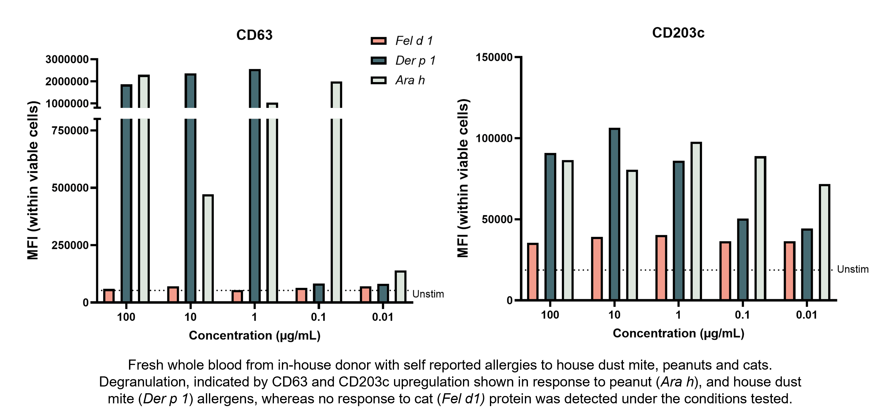
Degranulation is a hallmark of the immediate allergic response, occurring when allergen-mediated crosslinking of IgE-bound FcεRI triggers mast cell or basophil activation. Rapid release of preformed mediators, including histamine, proteases and β-hexosaminidase, contributes to the clinical manifestations of allergic inflammation, while subsequent production of lipid mediators and cytokines can sustain and amplify the response.
RoukenBio’s degranulation assays provide functional measures of IgE/FcεRI-mediated activation and therapeutic inhibition. Using primary or differentiated mast cells, as well as engineered reporter systems, we can quantify mediator release through endpoints including β-hexosaminidase, histamine, tryptase and surface activation markers such as CD63. These assays enable assessment of compound potency, mechanism of action and inhibition of mast cell or basophil effector function.

The Basophil Activation Test (BAT) provides a human primary-cell model of allergen-induced basophil activation. Allergen mediated crosslinking of FcεRI-bound IgE triggers basophil activation, resulting in rapid changes in surface activation markers and release of inflammatory mediators. As a circulating effector cell, the basophil provides a readily accessible model for investigating IgE dependent allergic responses in donor derived blood.
RoukenBio’s BAT platform uses flow cytometry to quantify basophil activation following allergen or receptor stimulation, with markers such as CD63 and CD203c providing sensitive measures of cellular activation. The assay can be applied to evaluate inhibitors of IgE/FcεRI signalling, assess mechanism of action and characterise inter-donor variability in allergic responses, providing a translational bridge between mechanistic assays and human biology.
Engineered reporter cell lines provide robust, sensitive and scalable platforms for the functional interrogation of signalling pathways implicated in allergic and type 2 inflammation. By coupling receptor activation to a quantifiable reporter response, these systems enable sensitive measurement of pathway activity and therapeutic inhibition with high assay consistency and throughput.
RoukenBio has developed a suite of reporter systems targeting key pathways in allergy, including TSLP/TSLPR, IL-4/IL-13/STAT6 and IgE/FcεRI signalling. Our NFAT-based reporter systems enable functional assessment of TSLP receptor and FcεRI activation, providing quantitative measures of receptor mediated signalling. Complementary IL-4/IL-13 reporter systems enable assessment of cytokine-induced STAT6 pathway activation. Together, these platforms support compound screening, potency assessment and mechanism-of-action studies, with orthogonal validation in primary human-cell assays to provide translational confidence in therapeutic activity across key pathways driving allergic and type 2 inflammation.

RoukenBio’s donor-derived Air–Liquid Interface (ALI) model provides a physiologically relevant in vitro system for investigating Type 2 airway inflammation and therapeutic responses. Primary human bronchial epithelial cells differentiate into a mucociliary epithelium containing ciliated and mucus-producing goblet cells.
IL-4/IL-13 stimulation induces disease-relevant features including mucus production, epithelial remodelling and inflammatory biomarker responses, enabling pharmacological activity to be assessed using complementary imaging and soluble endpoints.
Applications: Evaluation of Type 2 pathway inhibitors using functional and translational endpoints to support MoA studies and compound prioritisation.
Allergy programmes succeed when assays reflect human disease mechanisms and deliver reproducible, interpretable data. RoukenBio provides allergy therapeutic development services that combine translational immunology expertise with advanced primary cell work and engineered cellular tools, so you can progress faster through allergy drug discovery and candidate selection.
From the primary mast cell differentiation to functional assays, our in-house experts and novel solutions are here to guide you through the drug development pipeline.
We leverage our in-depth knowledge of immune cell function to provide reliable and reproducible results.
Tailored solutions to meet specific research needs in allergy and IgE-targeted therapy development.
Access to advanced flow cytometry, functional bioassays, and reporter-based systems.
Accelerate your allergy research and therapeutic development by contact us today to discuss how our deep immunology assay expertise can support your next breakthrough in allergy treatment.


An allergy assay is a laboratory model used to investigate immune mechanisms involved in allergic and type 2 inflammatory responses. Depending on the biological question, assays can assess IgE/FcεRI signalling, mast-cell and basophil activation, eosinophil function, type 2 cytokine signalling, epithelial responses and other disease-relevant pathways.
These assays can support target validation, potency assessment, mechanism-of-action studies, candidate selection and translational research.
Visual resources: Selected scientific illustrations on this website were created with BioRender.com and incorporate licensed BioRender content.
