
Dr Emilio Cosimo
Scientific Director of Innovation

 Insights
|
June 16, 2026
|
9 min read
FcRn biology is often reduced to a binding question: does a molecule bind FcRn at acidic pH and release at neutral pH? That question is important, but it is not the full biological story.
For antibodies and other Fc-containing biologics, FcRn-mediated recycling is a cellular rescue pathway. After cellular uptake, IgG can enter acidic endosomal compartments, bind FcRn, avoid lysosomal degradation, and return to the extracellular environment. A functional FcRn-mediated recycling assay is designed to measure the net outcome of that process, not binding alone. [1-4]
This distinction is key because FcRn binding affinity does not necessarily predict cellular recycling behaviour. Functional recycling data can add a biologically relevant layer to candidate evaluation, especially when interpreted alongside FcRn binding kinetics, developability profiling, internalisation data, and translational PK modelling. [5-7]
This article explains what FcRn-mediated recycling assays measure, how the data should be interpreted, and where these assays can support biologics development decisions.
FcRn is a salvage receptor for IgG and albumin. For IgG, its key role is to rescue a fraction of internalised antibody from intracellular degradation (Figure 1). [1-4]
Cells continuously internalise extracellular proteins through non-specific uptake pathways. In acidified endosomes, IgG can bind FcRn through its Fc region. FcRn-bound IgG is then sorted away from lysosomal degradation and returned to the cell surface. At near-neutral extracellular pH, the FcRn-IgG interaction weakens, allowing IgG release. [1-4]
This capture-and-release mechanism contributes to the long systemic persistence of many IgG molecules. For therapeutic antibodies and other Fc-containing biologics, systemic exposure can influence dosing interval, target coverage, therapeutic window, and the likelihood of achieving the intended pharmacological effect. [2-4,8-10]
FcRn is not the only determinant of pharmacokinetics. Target-mediated drug disposition, internalisation, antigen sink, non-specific binding, charge, hydrophobicity, aggregation, glycosylation, immunogenicity, and molecular format can all affect observed PK. FcRn should therefore be understood as one major component of antibody disposition, not the whole explanation. [8-12]
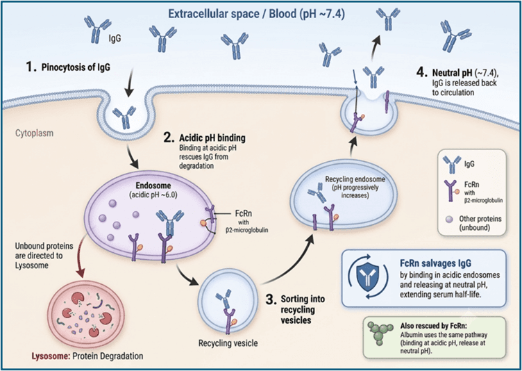
Figure 1. Mechanistic overview of FcRn-mediated recycling in cells. Following cellular uptake, IgG enters acidic endosomal compartments where FcRn binding can protect it from lysosomal degradation. The FcRn–IgG complex is then trafficked through the recycling pathway and returned to the cell surface, where neutral extracellular pH promotes dissociation and release. This diagram illustrates the underlying cellular mechanism that supports FcRn-mediated IgG salvage and recycling.
The most useful mental model for FcRn biology is not “stronger binding is better.” It is acidic-pH capture with efficient neutral-pH release. [5-7,13]
A molecule may benefit from sufficient FcRn binding in the acidic endosomal environment, typically around pH 6.0–6.5. However, measurable FcRn binding at physiological pH, or very slow release at neutral pH, can impair recycling and may worsen persistence rather than improve it. [5-7,13]
This is especially relevant for Fc engineering. Fc variants intended to extend half-life should be evaluated not only for acidic-pH binding, but also for neutral-pH release kinetics, whole-molecule developability, FcγR and C1q biology, and molecule-specific clearance risks. [14-16]
The FcRn-mediated recycling assay measures whether an IgG-based molecule or Fc-containing format can undergo a cellular FcRn-dependent rescue-and-release process. Conceptually, the assay asks three questions:
· Can the molecule access and engage the FcRn recycling pathway?
· Is the molecule recovered extracellularly after cellular handling?
· Is the observed recovery consistent with FcRn-dependent recycling rather than non-specific uptake, retention, or detection artefact?
A typical RoukenBio FcRn-mediated recycling assay design compares HEK293 WT cells with HEK293 FcRn-overexpressing cells (FcRn-OE). The WT condition helps define background recovery, while the FcRn-OE condition enriches the FcRn-dependent component of the assay response.
In a representative workflow (Figure 2), the test molecule is incubated with cells under assay-defined loading conditions. The assay then enables FcRn-dependent handling and extracellular release. Recovered IgG is measured in the supernatant, typically using ELISA or a related immunoassay format. Cell-associated material can also be measured in lysate to assess intracellular or retained signal.
The important point is that the assay does not simply measure FcRn binding. It measures a functional cellular outcome involving uptake, FcRn-dependent intracellular handling, protection from degradation, retention or release, and extracellular recovery.
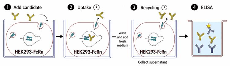
Figure 2. Representative workflow for the FcRn-mediated recycling assay. The candidate molecule is added to the cells and allowed to undergo uptake under assay-defined conditions. Following intracellular exposure, Fc-containing molecules that engage FcRn may be protected from degradation and recycled back toward the cell surface. After washing and addition of fresh medium, recovered IgG is released into the supernatant and quantified using ELISA or a related immunoassay format. Depending on assay design, this workflow can be performed under pH 6 or pH 7 conditions. This workflow provides a functional cellular readout of uptake, FcRn-dependent intracellular handling, and extracellular recovery, rather than a binding measurement alone.
In early discovery, FcRn recycling data can help compare candidates that look similar by target binding, potency, or FcRn binding affinity. A candidate with weaker functional recycling may carry additional uncertainty relevant to PK behaviour, especially if other developability liabilities are present. [5-7,12]
At this stage, the assay is most useful as a comparative tool. It can identify outliers, support ranking under matched conditions, and indicate whether further Fc engineering or developability work is justified.
For Fc-engineered molecules, the assay can support comparison of variants designed to alter FcRn interaction. This is particularly relevant for mutations intended to extend half-life by improving acidic-pH FcRn engagement while preserving neutral-pH release. [13-16]
The assay can help answer whether an engineered Fc behaves as intended in a cellular context. This is important because binding assays alone may not capture the combined effects of uptake, trafficking, release, and whole-molecule behaviour.
However, FcRn optimisation should not be treated as a default solution to every PK problem. If target-mediated clearance, antigen sink, non-specific tissue binding, aggregation, or poor developability dominates disposition, stronger FcRn interaction may not produce a meaningful exposure benefit. [8-12]
FcRn recycling is relevant to Fc-containing Fc-fusion proteins, bispecifics, ADCs, and other modified antibody formats, but interpretation must be modality-specific. [17-20]
For Fc-fusion proteins, the Fc can provide access to FcRn salvage, but the fusion partner may strongly influence uptake, intracellular retention, tissue distribution, target sink, and overall clearance. Fc-fusions should not be treated as smaller antibodies. [17,18]
For IgG-like bispecifics, FcRn salvage may be preserved, but altered architecture, charge distribution, stability, and self-association can affect PK. Fc-free bispecific formats lack canonical FcRn salvage and require different half-life-extension strategies. [17]
For ADCs, the intact antibody component may retain FcRn binding, but overall PK and developability can be dominated by drug-to-antibody ratio, hydrophobicity, linker stability, conjugation site, aggregation, target-mediated uptake, and deconjugation. FcRn recycling data should therefore sit alongside ADC-specific analytics rather than replace them. [19,20]
FcRn recycling data may also support CMC-relevant characterisation when FcRn-mediated handling is part of the molecule’s intended quality or developability profile. [21]
For example, the assay may help compare process variants, molecule versions, conjugation formats, or Fc-engineered versions where FcRn function is relevant to the product’s mechanism or value proposition.
This should be framed carefully. FcRn recycling data may contribute to a comparability-style or characterisation package, but it should not be presented as a standalone regulatory acceptance criterion. [21]
The same assay biology can support evaluation of investigational FcRn inhibitors. [22,23]
In this use-case, the intended pharmacology is reversed. Instead of supporting IgG rescue, an FcRn inhibitor is expected to reduce FcRn-mediated recycling and promote IgG degradation.
By measuring reduction of recovered IgG signal in FcRn-expressing cells, the assay can compare investigational inhibitors against reference FcRn inhibitors, including approved reference molecules such as Efgartigimod where scientifically justified and practically available. [22,23,26]
This application should be described as comparative in vitro pharmacology. Reduced recycling signal in vitro can support mechanism-consistent FcRn blockade, but it should not be interpreted as a standalone predictor of clinical IgG lowering, dose response, autoimmune disease efficacy, or regulatory acceptability. [22,23]
Functional FcRn recycling data can support translational PK assessment when combined with orthogonal evidence [8-12,24,25]. The most defensible interpretation comes from integrating:
· FcRn binding kinetics and affinity;
· neutral-pH release behaviour;
· cell-based recycling or transcytosis data;
· target binding and internalisation;
· receptor turnover and TMDD parameters;
· non-specific binding and developability profiling;
· species-aware in vivo PK data where relevant;
· mechanistic PK or PBPK modelling.
The assay can reduce uncertainty around FcRn-related behaviour. It cannot independently predict human half-life. [8-12,24,25]
FcRn-mediated recycling data should generally be interpreted qualitatively or comparatively unless quantitative thresholds have been established for the specific assay format, molecule class, and decision context.
A stronger recovered signal in FcRn-OE cells compared with WT cells may support an FcRn-dependent component of recycling (Figure 3). Reduced signal in the presence of an FcRn inhibitor supports pathway specificity. Differences between molecules may reflect Fc sequence, molecular format, conjugation chemistry, physicochemical properties, intracellular handling, or detection compatibility. [6,12]
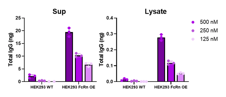
Figure 3. FcRn-overexpressing cells show increased recovered IgG compared with cells negative for FcRn. Representative FcRn-mediated recycling assay data, generated at pH 7, showing total IgG (Rituximab) recovered from supernatant and lysate fractions across candidate concentrations. FcRn-overexpressing cells show higher IgG recovery than cells negative for FcRn, supporting an FcRn-dependent component of the assay signal. The dose-dependent response in both supernatant and lysate fractions illustrates the importance of measuring released and cell-associated material when interpreting recycling efficiency.
Comparator molecules can help anchor interpretation. For example:
· an IgG antibody comparator can provide a reference recycling profile;
· an Fc-fusion comparator can contextualise format-dependent behaviour;
· an FcRn inhibitor comparator can support interpretation of pathway blockade.
The key point is not that any comparator is universal. Comparators are useful when they are run under matched assay conditions and interpreted within a defined decision context.
For FcRn inhibitor programs, interpretation is inverted. Reduced recycling signal may represent the desired pharmacological effect. In that setting, Efgartigimod can serve as an approved reference comparator for assessing whether a novel inhibitor produces a mechanism-consistent cellular inhibition profile. [22,23]
Higher recycling is therefore not always “better.” The optimal result depends on the product concept. For a half-life-extended antibody, efficient recycling may be desirable. For an FcRn inhibitor, reduced recycling is the intended effect. For some immunomodulatory or high-risk molecules, shorter persistence may be preferable. [5-7,13,22,23]
Robust FcRn recycling assays depend on appropriate controls and matched experimental conditions [5-7,12]. Important controls include:
· HEK293 WT versus FcRn-OE cells to distinguish FcRn-dependent signal from background.
· pH-dependent assay conditions that reflect the capture-and-release biology being tested.
· FcRn inhibition controls to support pathway specificity.
· Supernatant and lysate readouts to distinguish released material from cell-associated material.
· Reference molecules to support within-study ranking.
· Detection reagent controls to confirm that signal differences are not caused by assay interference.
· Cell health and expression controls to exclude artefacts from viability, receptor expression, or altered cell state.
Common pitfalls include:
· treating FcRn affinity as equivalent to functional recycling;
· using a single KD value without considering association and dissociation kinetics;
· overlooking neutral-pH binding or slow neutral-pH release;
· over-interpreting a single concentration or time point;
· ignoring lysate or cell-associated signal;
· comparing molecules across non-matched assay conditions;
· overlooking non-specific uptake, retention, aggregation, or detection interference;
· treating in vitro recycling differences as direct evidence of clinical PK or efficacy.
For FcRn inhibitor evaluation (Figure 4), an additional pitfall is equating cellular inhibition with clinical efficacy. Reduced recycling in vitro may support mechanism-consistent pharmacology, but clinical IgG lowering, disease-relevant pharmacodynamics, safety, and therapeutic effect require additional translational and clinical evidence. [22,23]
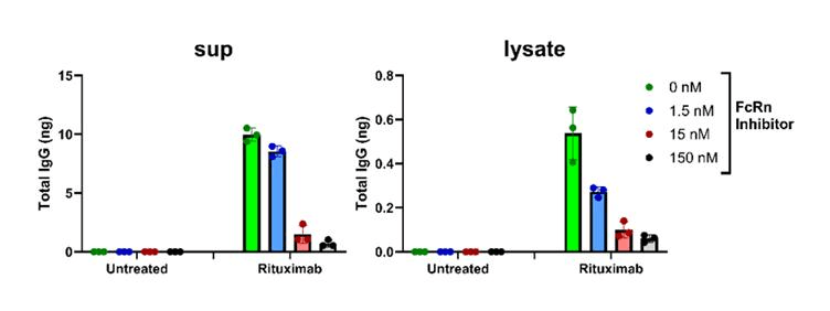
Figure 4. FcRn inhibition reduces recovered IgG signal in a cellular recycling assay. Representative cellular recycling data, generated at pH 7 in HEK293 FcRn-OE cells, showing reduced IgG recovery in the presence of increasing FcRn inhibitor concentrations. Reduced signal in both supernatant and lysate fractions is consistent with inhibition of FcRn-mediated IgG rescue and recycling under the assay conditions. This type of result can support comparative in vitro pharmacology for FcRn inhibitor candidates but should not be interpreted as a standalone predictor of clinical IgG lowering, dose response, or efficacy.
An FcRn-mediated recycling assay should not be interpreted as a direct predictor of human antibody half-life. [8-12,24,25]
FcRn biology is important, but PK is multi-factorial. A molecule with favourable recycling may still show poor PK if it has high target-mediated clearance, high non-specific binding, aggregation risk, poor stability, tissue retention, immunogenicity, or modality-specific clearance mechanisms. [8-12]
Conversely, a molecule with less favourable recycling may still be developable if its dosing strategy, therapeutic window, and efficacy profile support the intended use.
The FcRn-mediated recycling assay also does not replace:
· in vivo PK studies;
· translational PK/PD modelling;
· broader developability assessment;
· CMC comparability packages;
· clinical pharmacology;
· pharmacodynamic assessment of IgG lowering for FcRn inhibitors;
· disease-relevant efficacy studies.
Its value is strongest when used to reduce uncertainty, compare candidates under matched conditions, and identify mechanistic risks early enough to inform design or prioritisation decisions.
Functional FcRn recycling data is most informative when interpreted alongside orthogonal evidence. [5-7,12]
SPR-based FcRn binding analysis is a useful complementary method because it quantifies binding kinetics and affinity under defined pH conditions. These measurements help show whether an antibody, Fc-fusion protein, ADC, or FcRn inhibitor engages FcRn as intended. [5,7]
However, SPR and cellular recycling assays answer different questions.
SPR measures molecular interaction under controlled biophysical conditions. The FcRn-mediated recycling assay measures the cellular outcome of uptake, receptor engagement, intracellular handling, retention, and release. [5-7,12]
This distinction is critical. Two antibodies may show broadly similar apparent FcRn binding by SPR under acidic conditions yet display different cellular recycling profiles (Figure 5). That is not contradictory. FcRn binding is one component of recycling, but cellular recovery also reflects trafficking, release efficiency, non-specific handling, molecule format, and detection factors. [5-7,12,26]
The practical implication is that SPR and functional recycling assays should not be treated as interchangeable. Binding data establishes whether FcRn engagement occurs under defined conditions. Functional recycling data adds a cellular layer that can reveal differences not apparent from binding affinity alone. [5-7,12]
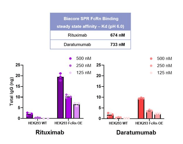
Figure 5. Similar FcRn binding by SPR does not necessarily imply equivalent cellular recycling. Representative orthogonal data comparing FcRn binding and cellular recycling for Rituximab and Daratumumab under their respective assay conditions. SPR analysis, performed at pH 6, shows broadly similar apparent FcRn binding affinity for the two antibodies, with steady-state KD values in the sub-micromolar range. In contrast, the cellular recycling assay, performed at pH 7, shows distinct recovered IgG profiles in HEK293 WT cells (negative for FcRn) and HEK293 FcRn-OE cells. These data illustrate why FcRn binding assays and FcRn-mediated recycling assays should be interpreted as complementary readouts: SPR confirms molecular interaction with FcRn, while the cellular assay captures the net outcome of uptake, FcRn-dependent intracellular handling, and extracellular recovery.
Relevant supporting assays may include:
· FcRn binding kinetics and affinity analysis by SPR, BLI, or related biophysical methods.
· Solution competition assays or FcRn chromatography to test pH-dependence and reduce surface artefacts.
· Target binding and potency assays.
· Internalisation and receptor turnover assays for TMDD-relevant targets.
· Fcγ receptor and C1q binding assays where effector biology may matter.
· Aggregation, stability, hydrophobicity, charge, self-interaction, and broader developability profiling.
· ADC-specific analytics including DAR distribution, linker stability, HIC/SEC, and deconjugation assessment.
· In vitro FcRn inhibitor benchmarking against appropriate reference molecules.
· Species-aware in vivo PK studies where scientifically justified.
· Mechanistic PK or PBPK modelling where sufficient supporting data are available.
Together, these assays provide a more complete view of antibody disposition or FcRn inhibitor pharmacology than any single method can provide. [5-12,24,25]
FcRn recycling data can help compare Fc variants, identify unintended effects of engineering, and support rational selection of molecules with appropriate pH-dependent behaviour. [13-16]
The assay is most useful when FcRn biology is part of the design strategy and when results are interpreted alongside neutral-pH release, FcγR/C1q biology, and developability data.
The assay provides a functional cellular readout that complements biophysical binding methods. [5-7,12]
Its value depends on robust controls, matched conditions, appropriate detection reagents, cell-state monitoring, and clear separation of extracellular recovered material from intracellular or cell-associated material.
The assay can support comparative evaluation of FcRn inhibitors by measuring functional blockade of IgG recycling in cells. [22,23]
Reference inhibitors such as Efgartigimod may help benchmark cellular inhibition profiles. These data can support early pharmacology and assay confidence, but they should not be overextended into clinical efficacy claims. [22,23]
For program decision-makers, the assay helps translate FcRn biology into development risk language.
It can support candidate comparison, identify FcRn-related uncertainty, and clarify whether additional Fc engineering, developability work, CMC-supportive characterisation, inhibitor benchmarking, or translational modelling is justified before larger resource commitments.
Myth 1: FcRn binding alone explains antibody half-life.
Reality: FcRn binding is important, but half-life is influenced by target biology, TMDD, non-specific uptake, developability, immunogenicity, molecular format, and species translation. [8-12]
Myth 2: Higher FcRn affinity is always better.
Reality: Higher acidic-pH affinity may be useful only if neutral-pH release remains efficient. Neutral-pH binding or slow release can impair recycling. [5-7,13]
Myth 3: Higher FcRn recycling is always desirable.
Reality: The desired recycling profile depends on the product concept. For FcRn inhibitors, reduced recycling is the intended pharmacological effect.
Myth 4: FcRn recycling assays directly predict human PK.
Reality: FcRn recycling assays provide mechanistic in vitro data. They support interpretation and candidate comparison but do not replace in vivo PK, translational modelling, or clinical data. [8-12,24,25]
FcRn-mediated recycling assays are best viewed as mechanistic decision tools. They do not provide a standalone answer to PK, but they can help clarify whether Fc-containing molecules behave as expected in a cellular FcRn-dependent system.
Used appropriately, these assays can support candidate selection, Fc engineering, developability assessment, CMC-supportive characterisation, translational PK assessment, and FcRn inhibitor benchmarking.
For antibodies, Fc-fusion proteins, ADCs, Fc-engineered molecules, and FcRn inhibitors, functional recycling data can add useful biological context before costly downstream decisions are made.
🗓️ Stay informed with our monthly scientific newsletter, published on LinkedIn on the last Wednesday of each month.
These editions bring you the latest in drug development breakthroughs, industry trends, and expert insights from the brilliant minds at RoukenBio.
Subscribe today on LinkedInRoukenBio’s FcRn-mediated recycling assay is designed to generate functional FcRn recycling data that complements FcRn binding, developability, and translational PK assessment workflows.


 Research Posters