Developing effective asthma therapeutics requires disease models that represent the structure and responsiveness of the human airway. RoukenBio’s donor-derived air-liquid interface (ALI) asthma model recreates key epithelial features associated with Type 2 airway inflammation and generates measurable endpoints for therapeutic evaluation, target validation and mechanism-of-action studies. Quantify therapeutic activity in a differentiated human donor airway model with greater translational confidence.

Asthma affects hundreds of millions of people worldwide, yet it is far from a single disease. Researchers now recognise multiple asthma endotypes, including Type 2 (Th2)-driven, neutrophilic and paucigranulocytic asthma, each characterised by distinct underlying biological mechanisms. This complexity means that therapies that are effective for one patient population may not show the same efficacy in another, creating significant challenges for target validation, biomarker discovery and therapeutic development. Supporting evidence from the wider scientific literature also highlights asthma's molecular and clinical heterogeneity as a major obstacle to developing universally effective treatments.
Adding to this challenge, many conventional preclinical models do not fully replicate human airway biology. Conventional submerged cultures do not reproduce the differentiated structure of the airway epithelium while animal models may not accurately reflect human-specific physiology, immune responses or disease mechanisms. As a result, promising therapeutic candidates can be difficult to evaluate using models that do not adequately capture clinically relevant disease features.

This has driven growing interest in human-relevant New Approach Methodologies (NAMs) and advanced in vitro systems capable of bridging the gap between simple laboratory assays and clinical studies. Donor-derived Air-Liquid Interface (ALI) models offer one such approach by recreating key features of the human airway microenvironment, including differentiated mucociliary epithelium, cytokine-driven epithelial remodelling, mucus hypersecretion and disease-relevant biomarker responses. By providing quantifiable and translationally relevant readouts, ALI models can help companies generate more meaningful insights earlier in the drug discovery process and support informed decision-making during therapeutic development.
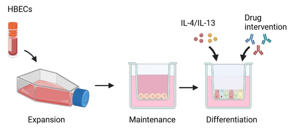
Primary human bronchial epithelial cells (HBECs) are expanded and seeded onto permeable inserts. Following a short maintenance phase, culture medium is supplied to the basal surface while the apical surface is exposed to air. Over a differentiation period of approximately three to four weeks, the cells develop into a three-dimensional pseudostratified mucociliary epithelium containing specialised airway cell populations, including goblet cells and ciliated cells.
Stimulation with IL-4 and IL-13 enables controlled induction of asthma-like phenotypes ready for targeted drug intervention. Models can be generated using cells from healthy donors or asthmatic patients.
Primary human bronchial epithelial cells are differentiated under ALI conditions to form a three-dimensional pseudostratified mucociliary epithelium. Stimulation with IL-4 and/or IL-13 induces asthma-associated epithelial changes, enabling test articles to be assessed against disease-relevant structural, functional and soluble readouts.
Donor-derived airway epithelium with differentiated goblet cells and ciliated cells.
Controlled induction of Type 2 asthma-associated biology with demonstrated pathway responsiveness.
Mucus production, soluble biomarker endpoints, confocal imaging, histology.
Airway Epithelial Remodelling
Mucus Hypersecretion
Biomarker Analysis
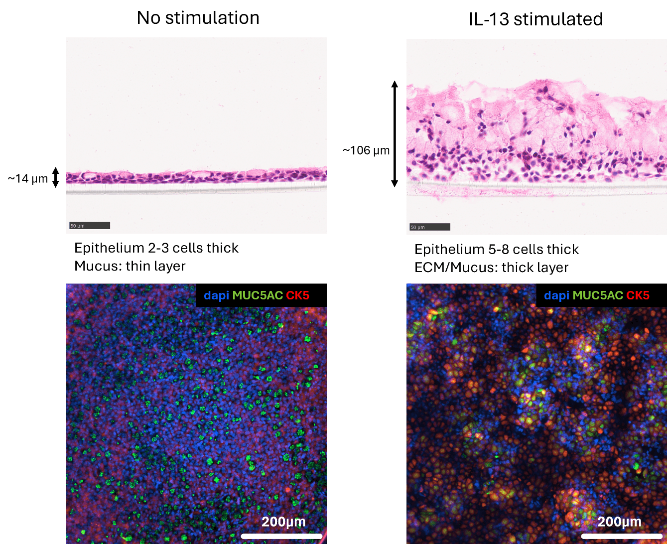
Following IL-13 stimulation, cultures display substantial structural changes consistent with airway remodelling observed in patients with asthma, including increased epithelial thickness, altered tissue organisation and accumulation of extracellular material and mucus.
Following IL-13 stimulation, cultures display substantial structural changes consistent with airway remodelling observed in patients with asthma, including increased epithelial thickness, altered tissue organisation and accumulation of extracellular material and mucus.
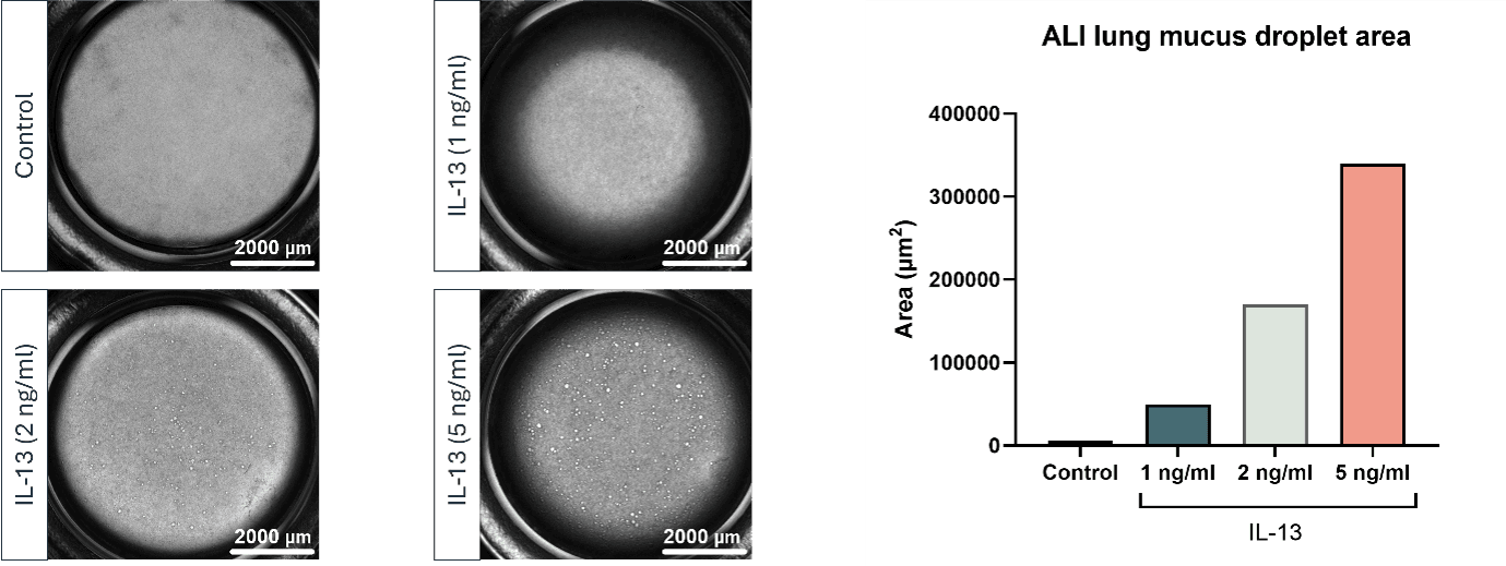
Excessive mucus production is a hallmark of Type 2 asthma. Within the ALI model, IL-13 stimulation drives concentration-dependent increases in mucus formation, generating a quantifiable assay readout for therapeutic evaluation.
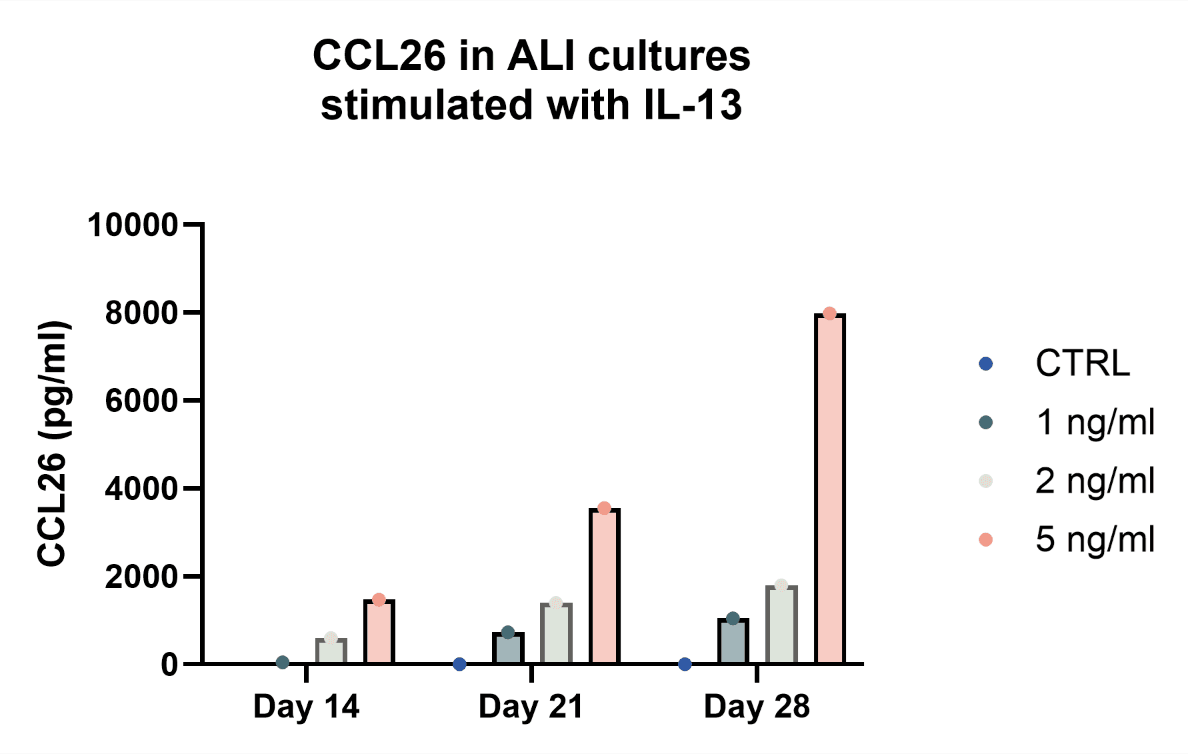
The platform supports analysis of disease-associated soluble mediators. In the development dataset, IL-13 stimulation produced concentration-dependent increases in CCL26 across multiple collection points. This adds a soluble biomarker endpoint to the imaging and histological assessment of the model.
The donor-derived ALI asthma model can support:
Generation of translational datasets to support compound prioritisation and programme progression.
RoukenBio is continuing the development of the platform towards a miniaturised 96-well HTS format to support broader donor profiling and higher-throughput compound evaluation. Ongoing development efforts include expansion of donor biobanks, refinement of imaging workflows and investigation of additional asthma subtypes and cell types relevant to asthma pathogenesis.
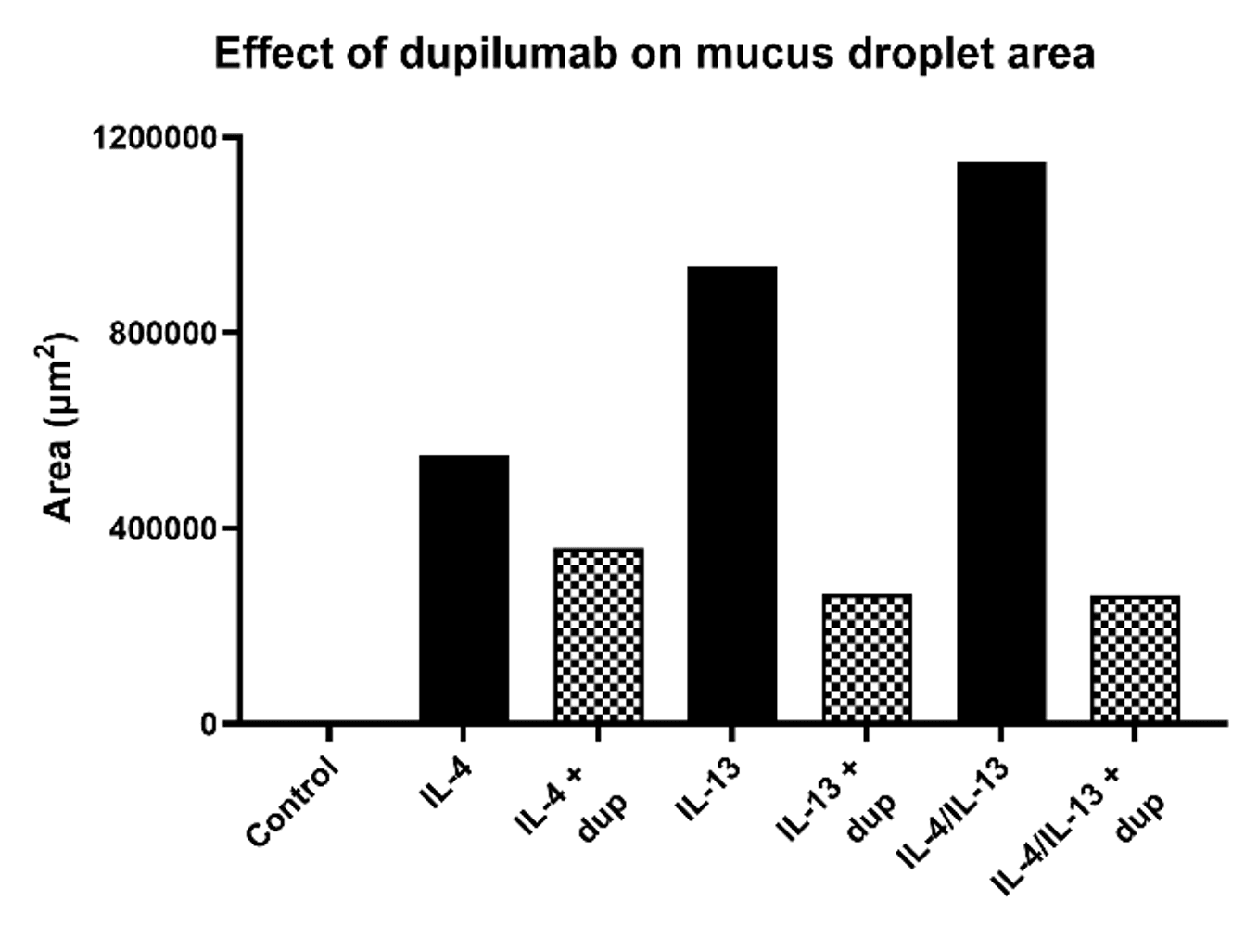
A drug-discovery model is most useful when a relevant intervention produces a measurable change in the induced phenotype. RoukenBio evaluated the platform using dupilumab, an IL-4Rα-blocking monoclonal antibody that inhibits IL-4 and IL-13 signalling.
In cytokine-stimulated cultures, dupilumab reduced mucus droplet formation across IL-4, IL-13 and combined IL-4/IL-13 conditions. This demonstrates the platform’s ability to detect a biologically meaningful response to pathway-targeted intervention and supports its use for therapeutic evaluation and mechanism-of-action studies.
Our model uses donor-derived human airway epithelial cells cultured at an air-liquid interface to more closely replicate the structure and function of the human airway than conventional submerged cultures.
Our air-liquid interface platform forms a mature mucociliary airway epithelium with specialised goblet and ciliated cells, enabling the study of human airway biology.
Following cytokine stimulation, the model exhibits several hallmark characteristics of Type 2 airway inflammation, including:
The platform incorporates multiple measurable endpoints to support therapeutic assessment, including:
Imaging-based readouts
The model responds to pathway-targeted intervention, supporting its use in therapeutic evaluation. Treatment with dupilumab reduced mucus droplet formation in cytokine-stimulated cultures, demonstrating the ability of the platform to detect biologically meaningful responses.
Access to healthy and asthma donor-derived airway cultures with flexible cytokine stimulation and study designs, enabling comparison across disease states and supporting therapeutic evaluation in diverse biological settings.
Discover how RoukenBio's donor-derived organotypic in vitro ALI asthma model can help generate translationally relevant data, evaluate therapeutic activity and provide quantifiable endpoints for respiratory drug discovery programmes.

An Air-Liquid Interface (ALI) asthma model is a donor-derived human airway epithelial culture system that recreates key features of the airway microenvironment. Primary human bronchial epithelial cells are cultured on permeable inserts, with culture medium supplied to the basal surface while the apical surface remains exposed to air. Over several weeks, the cells differentiate into a three-dimensional pseudostratified mucociliary epithelium containing goblet cells and ciliated cells. This enables researchers to study airway epithelial responses in a more physiologically relevant setting than conventional submerged cell cultures.
