Explore RoukenBio’s FcRn recycling assay for functional cell-based assessment of antibodies, Fc-fusions, ADCs and FcRn inhibitors. Generate FcRn ELISA readouts to support candidate selection, Fc engineering and translational PK assessment.

FcRn binding affinity alone does not always predict how an IgG-based biologic behaves in a cellular recycling pathway.
RoukenBio’s FcRn recycling assay provides a functional, cell-based readout of FcRn-mediated rescue and release, helping biologics teams evaluate antibodies, Fc-engineered molecules, Fc-fusion proteins, ADCs and FcRn inhibitors beyond binding measurements alone.
The assay measures the net cellular outcome of uptake, FcRn engagement, intracellular handling and extracellular recovery. This provides a more biologically relevant view of FcRn-mediated recycling than an FcRn binding assay or FcRn ELISA binding readout alone.

The neonatal Fc receptor, or FcRn, is a central regulator of IgG homeostasis and systemic persistence.
After cellular uptake, IgG can enter acidic endosomal compartments where FcRn binding protects a proportion of internalised antibody from lysosomal degradation. The FcRn-IgG complex can then recycle back to the cell surface, where near-neutral pH promotes antibody release.
This rescue-and-release process can influence systemic exposure, half-life, dosing strategy and developability risk for many Fc-containing biologics.
However, FcRn is not the only determinant of pharmacokinetics. Target-mediated drug disposition, internalisation, antigen sink, aggregation, charge, hydrophobicity, molecular format, tissue distribution and immunogenicity can all affect in vivo behaviour.
That is why FcRn recycling data should be interpreted as part of a broader developability and translational PK strategy.
Why Binding Data Alone Is Not Enough
FcRn Binding Assay vs FcRn Recycling Assay
FcRn binding assays, including SPR and BLI formats, are valuable tools for measuring molecular interaction with FcRn under defined conditions.
They help answer questions such as:
But FcRn-mediated recycling is not just a binding event.
A molecule must also access the relevant intracellular pathway, engage FcRn in the appropriate compartment, avoid degradation, traffic through recycling routes and release efficiently at neutral pH.
Two molecules may show similar FcRn binding affinity yet display different cellular recycling profiles. This is why a functional FcRn-mediated recycling assay can add important biological context to FcRn binding data.
FcRn binding assays, including SPR and BLI formats, are valuable tools for measuring molecular interaction with FcRn under defined conditions.
They help answer questions such as:
But FcRn-mediated recycling is not just a binding event.
A molecule must also access the relevant intracellular pathway, engage FcRn in the appropriate compartment, avoid degradation, traffic through recycling routes and release efficiently at neutral pH.
Two molecules may show similar FcRn binding affinity yet display different cellular recycling profiles. This is why a functional FcRn-mediated recycling assay can add important biological context to FcRn binding data.
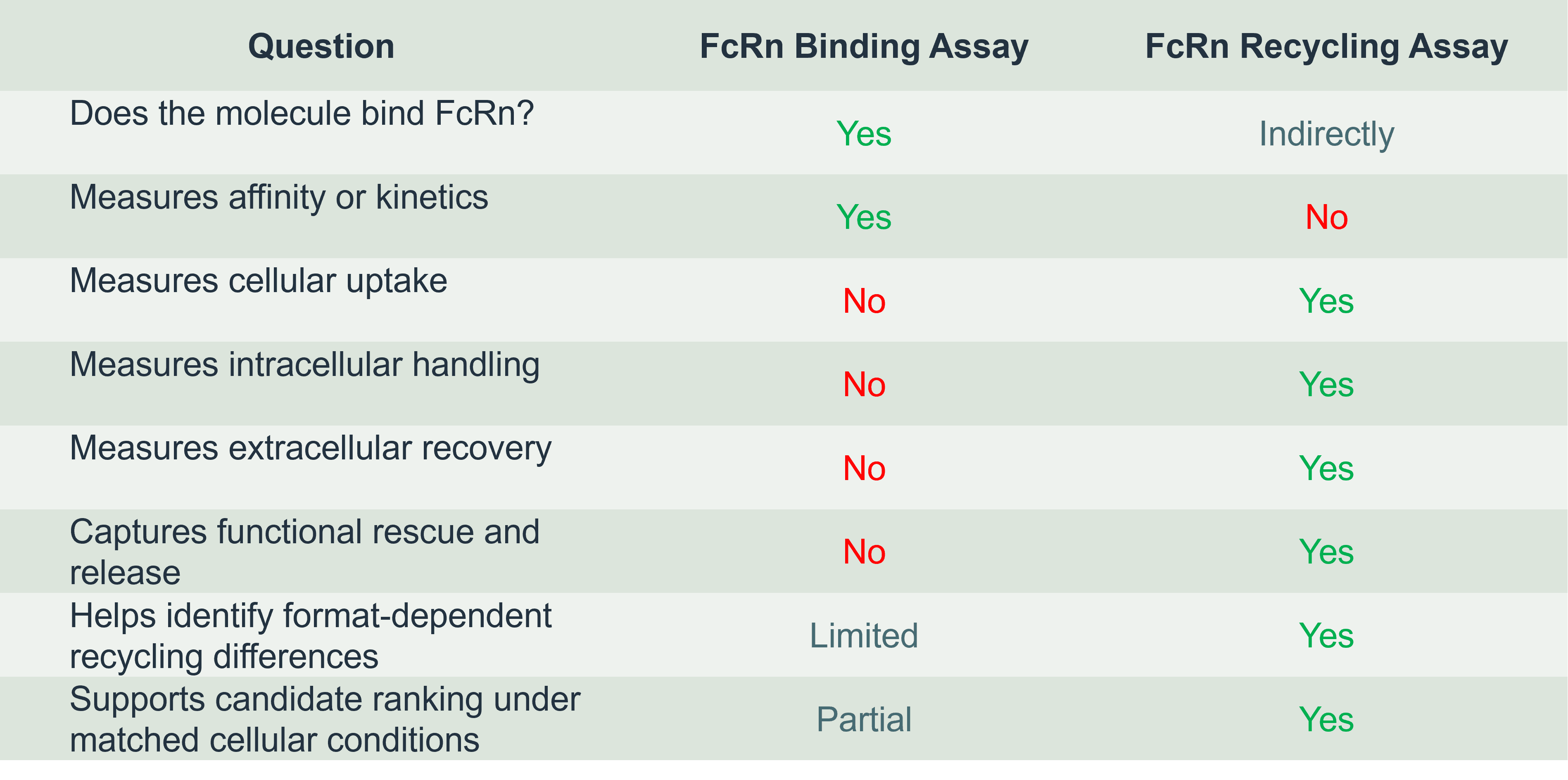
The two approaches are complementary. FcRn binding assays define receptor engagement. FcRn recycling assays measure the functional cellular outcome.
An FcRn recycling assay is a functional cell-based assay designed to assess whether an IgG-based molecule can undergo FcRn-mediated rescue from intracellular degradation and return to the extracellular compartment.
The assay is particularly useful when teams need to understand FcRn biology in a cellular context rather than relying only on equilibrium binding, SPR kinetics or FcRn ELISA binding data.
What the FcRn Recycling Assay Measures
Assay Format and FcRn ELISA Readouts
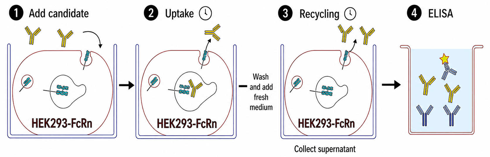
Figure 1. Schematic overview of the FcRn recycling assay workflow
The assay evaluates whether a molecule can productively engage the FcRn recycling pathway and be recovered after intracellular handling.
In practical terms, the assay addresses three development-relevant questions:
The assay assesses whether the candidate can enter the cellular environment and interact with FcRn-dependent recycling biology under controlled conditions.
Recycled material is measured in the supernatant, providing a readout of extracellular recovery following intracellular processing.
Control conditions help distinguish FcRn-mediated recycling from non-specific uptake, retention, degradation or detection artefact.
This is important because FcRn binding affinity alone does not necessarily predict functional recycling efficiency in cells.
Figure 1. Schematic overview of the FcRn recycling assay workflow
The assay evaluates whether a molecule can productively engage the FcRn recycling pathway and be recovered after intracellular handling.
In practical terms, the assay addresses three development-relevant questions:
The assay assesses whether the candidate can enter the cellular environment and interact with FcRn-dependent recycling biology under controlled conditions.
Recycled material is measured in the supernatant, providing a readout of extracellular recovery following intracellular processing.
Control conditions help distinguish FcRn-mediated recycling from non-specific uptake, retention, degradation or detection artefact.
This is important because FcRn binding affinity alone does not necessarily predict functional recycling efficiency in cells.
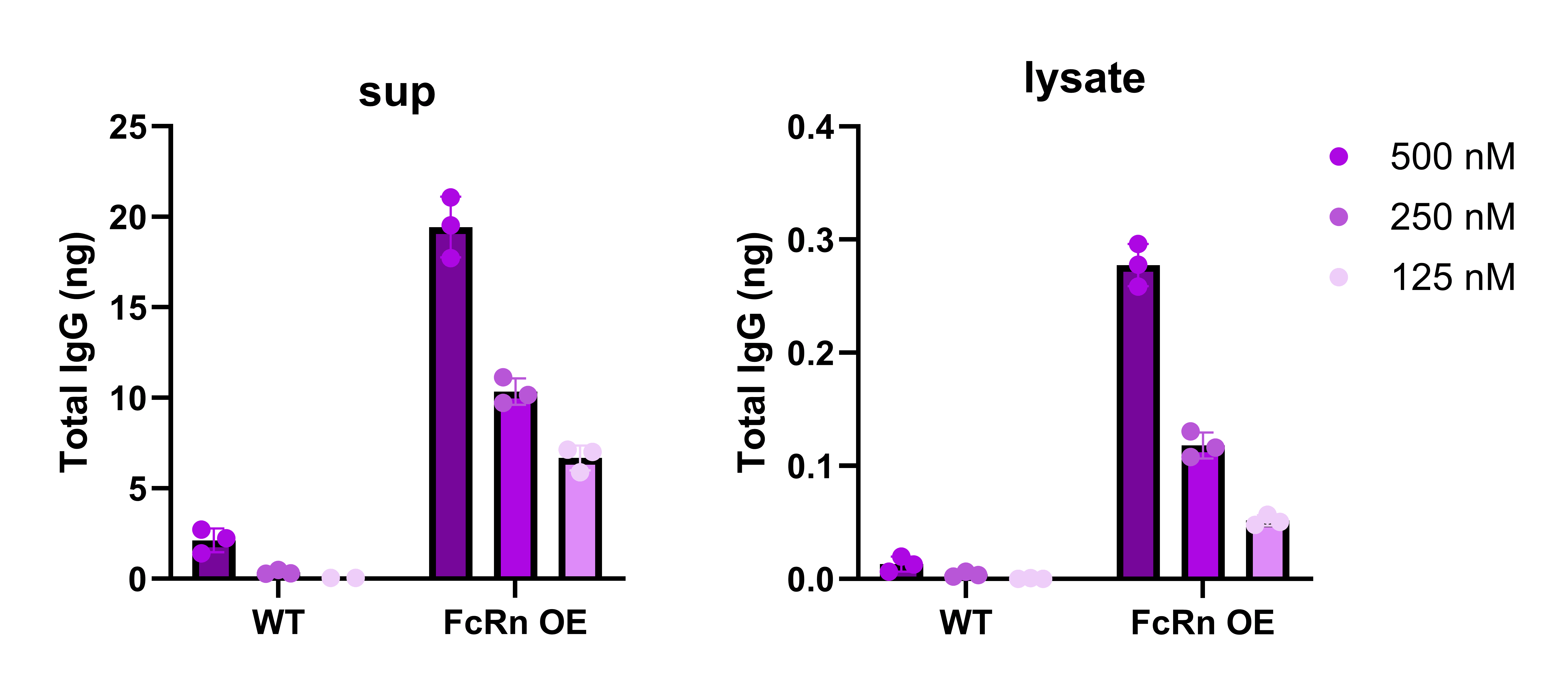
Figure 2. Rituximab recycling profile. Supernatant and intracellular lysate signal across increasing concentrations at pH 7.3. Clear separation from WT controls at all concentrations.
The assay uses HEK293 wild-type (WT) and FcRn-overexpressing (FcRn OE) cell systems to distinguish FcRn-dependent signal from background cellular handling.
Following molecule loading and intracellular exposure, recycled material is measured in the supernatant. Cell-associated material can also be measured in lysate, providing a fuller view of release versus residual.
Readout is typically performed using an ELISA-based detection format, enabling quantitative comparison across molecules, concentrations and experimental conditions.
Assay specificity can be supported through:
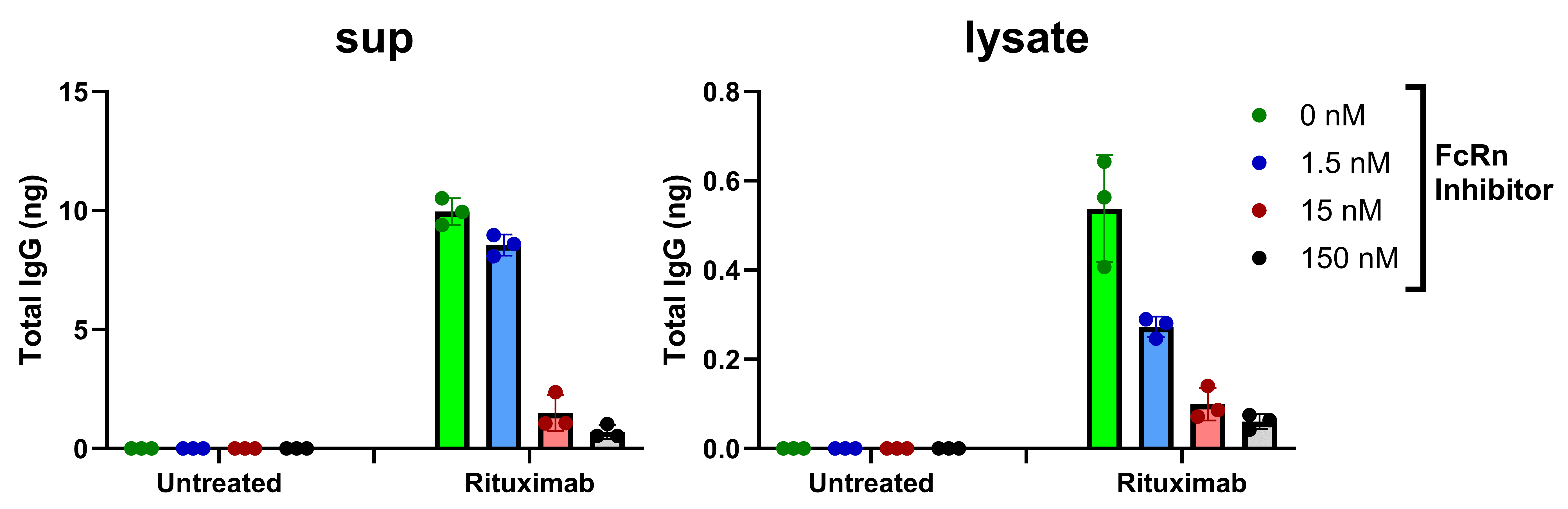
Figure 3. FcRn inhibitor effect on Rituximab. Recycling signals reduced to near-zero at 150 nM inhibitor in FcRn-OE cells at pH 7.3, confirming assay specificity.
Together, these controls help determine whether the observed signal is consistent with FcRn-mediated recycling rather than non-specific cellular behaviour.
Candidate Selection
Fc Engineering and Half-Life Optimisation
Fc-Fusion Proteins
Antibody-Drug Conjugates
FcRn Inhibitor Evaluation
Supporting Translational PK Assessment
In discovery and early lead selection, FcRn recycling data can help compare candidates under matched cellular conditions.
This is useful when molecules appear similar by target potency, FcRn binding affinity or developability profile but may behave differently in a functional recycling pathway.
The assay can help identify outliers, support ranking decisions and highlight candidates that may require further Fc engineering or developability assessment.
In discovery and early lead selection, FcRn recycling data can help compare candidates under matched cellular conditions.
This is useful when molecules appear similar by target potency, FcRn binding affinity or developability profile but may behave differently in a functional recycling pathway.
The assay can help identify outliers, support ranking decisions and highlight candidates that may require further Fc engineering or developability assessment.
Fc engineering strategies often aim to improve systemic exposure by modifying FcRn interaction.
However, stronger FcRn binding is not automatically better.
The most useful FcRn profile generally involves sufficient acidic-pH engagement with efficient release at near-neutral pH. Excessive neutral-pH binding or slow release may impair recycling rather than improve it.
A cell-based FcRn recycling assay can help assess whether Fc-engineered variants behave as intended in a functional cellular context.
Fc-fusion proteins can access FcRn salvage pathways through their Fc domain, but their behaviour is not identical to that of conventional IgG antibodies.
The fusion partner may influence uptake, intracellular retention, target-mediated clearance, tissue distribution and overall PK.
A functional FcRn recycling assay can support comparative profiling of Fc-fusion formats and help determine whether recycling behaviour is consistent with the intended developability profile.
For ADCs, the intact antibody component may retain FcRn interaction, but overall disposition can be influenced by conjugation-dependent properties.
Relevant factors include:
An ADC FcRn recycling assay can help compare whether different conjugation strategies alter FcRn-mediated recycling behaviour under matched conditions.
This should be interpreted alongside ADC-specific analytics such as DAR distribution, HIC, SEC, linker stability and deconjugation assessment.
The same assay biology can also support FcRn inhibitor programmes.
In this setting, the desired biological outcome is reduced FcRn-mediated IgG recycling.
An FcRn inhibitor assay can measure reduction in recovered IgG signal in FcRn-expressing cells, providing a functional cellular readout of FcRn blockade.
This can support comparative in vitro pharmacology and benchmarking against reference FcRn inhibitors where appropriate.
Reduced recycling in vitro should not be interpreted as a standalone predictor of clinical IgG lowering, dose response or efficacy. It is a mechanism-consistent cellular readout that should be interpreted alongside pharmacodynamic, translational and clinical evidence.
FcRn-mediated recycling is an important component of antibody disposition, but pharmacokinetics remains multifactorial.
Functional FcRn recycling data is most informative when integrated with orthogonal datasets, including:
Used in this context, FcRn recycling data can help reduce uncertainty around FcRn-related behaviour and support more informed decisions across discovery, lead optimisation and translational development.
Development Stages
Interpreting FcRn Recycling Data Correctly
Common Pitfalls in FcRn Assay Strategy
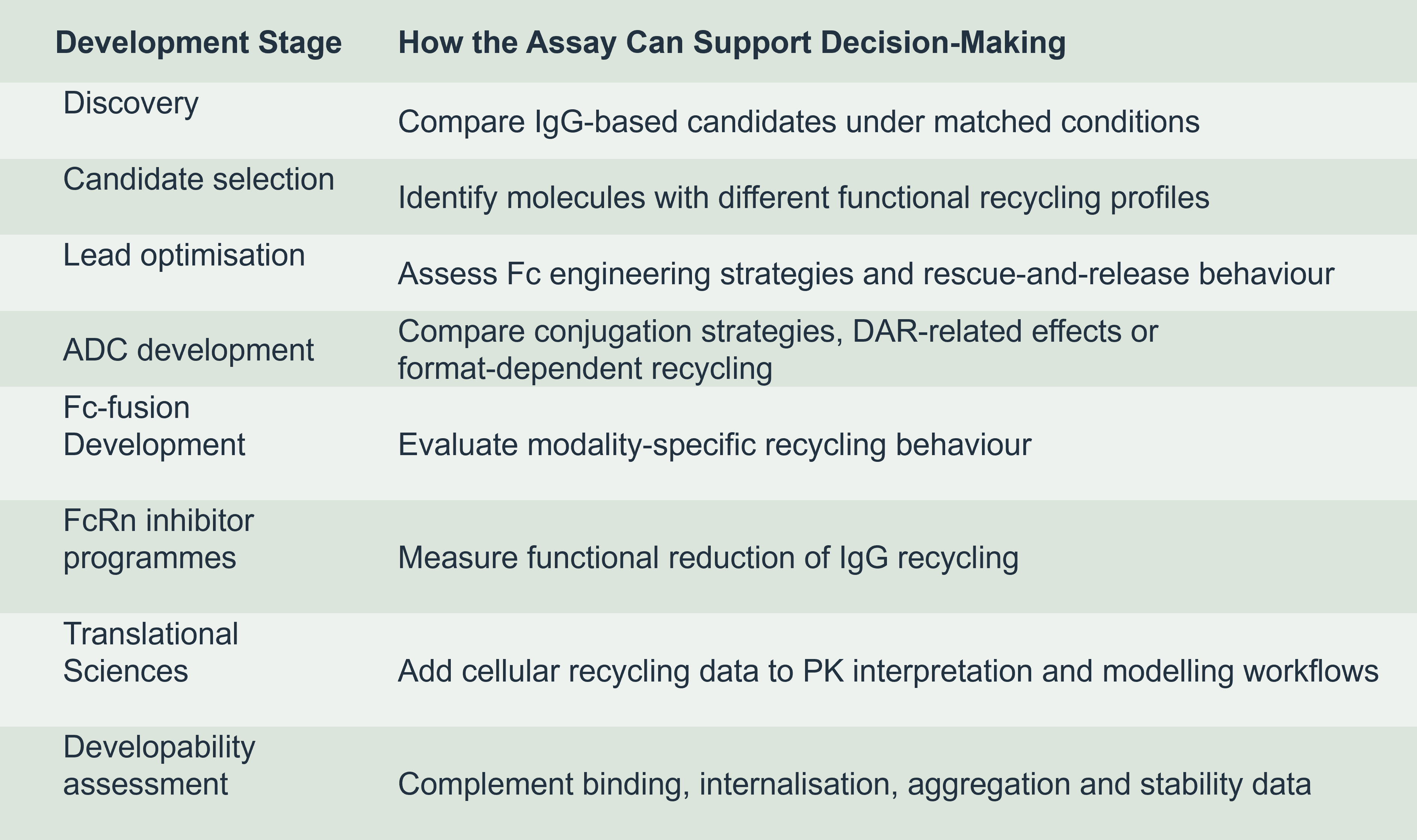
FcRn recycling data should generally be interpreted comparatively and in context.
A stronger recovered signal in FcRn-overexpressing cells compared with wild-type controls may support an FcRn-dependent component of recycling.
Reduced signal in the presence of an FcRn inhibitor can support pathway specificity.
Differences between molecules may reflect Fc sequence, molecular format, conjugation chemistry, physicochemical properties, intracellular handling or detection compatibility.
The strongest interpretation comes from combining FcRn recycling data with FcRn binding, developability, internalisation and translational PK evidence.
Treating FcRn affinity as equivalent to recycling
FcRn binding is necessary for recycling biology, but it is not the whole process. Functional recycling also depends on cellular uptake, trafficking, release, retention and degradation.
Assuming higher FcRn binding is always better
Improved acidic-pH binding may be useful only if neutral-pH release remains efficient.
Ignoring lysate or cell-associated signal
Supernatant data alone may not distinguish efficient release from intracellular retention. Lysate readouts can provide important context.
Comparing data across non-matched conditions
FcRn recycling data is most useful when molecules are compared under matched assay conditions.
We work with biologics teams that need more than a standard binding readout.
RoukenBio’s FcRn-mediated recycling assay is designed to generate functional cellular data that complements FcRn binding, developability and translational PK workflows.
This helps FcRn biology sit within the full development context rather than being treated as an isolated assay result.

Explore the biological rationale for FcRn-mediated recycling assays, assay workflow and experimental design with more data.

An FcRn recycling assay is a functional cell-based assay designed to assess whether an IgG-based molecule can undergo FcRn-mediated rescue from intracellular degradation and return to the extracellular compartment.
Rather than measuring binding alone, the assay captures the net cellular outcome of uptake, FcRn engagement, intracellular handling and extracellular recovery.
